FIELD VETERINARY REPORT FOR MASAI MARA –MARCH 2016 By
FIELD VETERINARY REPORT FOR MASAI MARA –MARCH 2016
By.Dr. Campaign K. Limo
Introduction
The Month was characterised by hot conditions during the day with very little to no precipitation. The mysterious deaths of elephants that occurred the previous month were not encountered during the month with results from the Government chemist indicating carbamate pesticides being detected in the stomach contents of the samples that were collected. There is still plenty of forage and water for the wildlife to utilize though.
The following are activities carried out by the unit during the month of March.
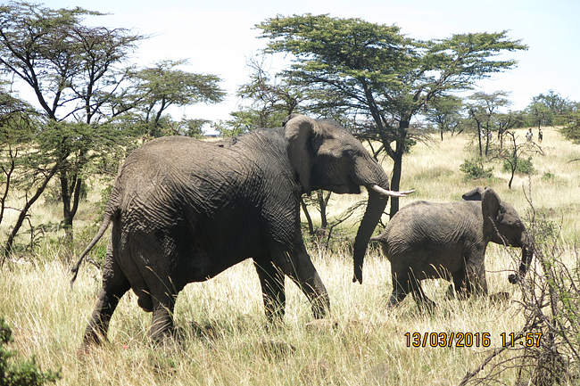
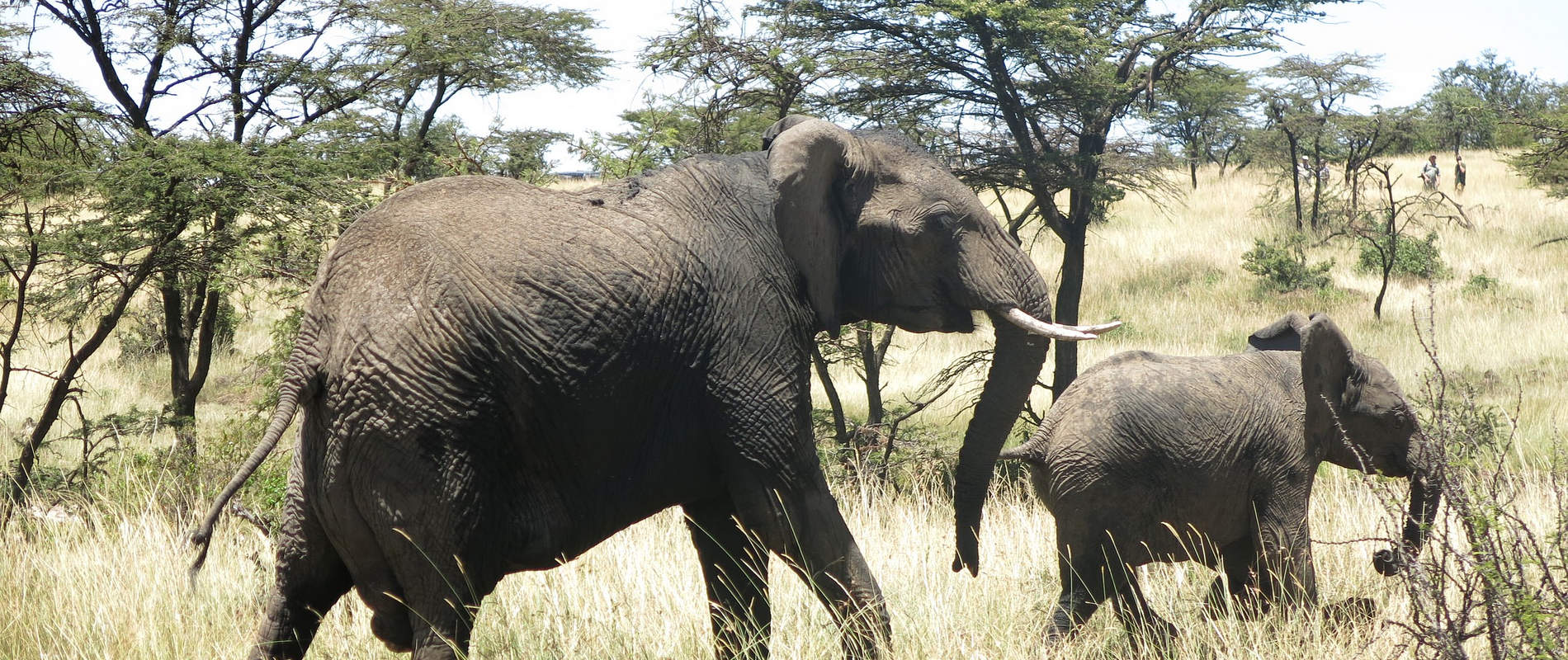
CASE#1 REPORT ON TWO DEAD ELEPHANTS
History
Reports were made to the mobile veterinary unit by MMNR that two elephant carcasses had been seen the morning of 6th March 2016 at GPS 36M 0735202 UTM 9838673 Olkeju Ronkai and 36M 0735631UTM 9843887 Olkiombo within MMNR. It was therefore necessary to confirm and determine the cause of their deaths.
Elephant 1
This elephant carcass at Olkeju Ronkai area in MMNR was found in a lugga estimated to have been more than ten days old. This was an approximately ten year old elephant whose sex could not be determined owing to the state of degradation. Part of the bony skeleton could only be seen. Both tusks were intact and recovered by MMNR security team for accounting and custody. The cause of death for this elephant could not be determined grossly owing to the state of decay and no meaningful samples to assist investigate its cause could be collected from the carcass.


Elephant 2
The carcass of this elephant was found the same day near Olkiombo airstrip about fifteen kilometres from the initial carcass. This was estimated to be 3 to 4 days old carcass of a young elephant whose sex could not be determined because the carcass had been extensively devoured by scavengers. Only the skull could be seen and a portion of hind limb which was partly submerged in a muddy pool. Hyenas and other scavengers had completely dismembered and stripped the carcass. As such no meaningful samples could be collected from it. Both tusks were intact and were recovered by MMNR for accounting and safe custody. Its cause of death could therefore not be determined.


Conclusion
Owing to the state of the two carcasses, their cause of death could not be determined. Moreover, no viable samples could be collected to assist investigating the cause of their deaths.
CASE#2 RE-TREATING A BULL ELEPHANT
Date: 7th March 2016
Species: African Elephant
Sex: Male
Age: Adult
Location: Mara Triangle Conservancy
GPS: 36M0720778 UTM 9852042
This bull had been treated in May 2015 for a severe wound created by a cable wire snare on his left foot. The snare was removed and the wound treated. Several tendons had been severed completely. Despite this, this bull went on to recover though the whole limb remained swollen. Earlier this year, this bull was treated through the sky vet initiative for a wound on his left foot pad. There was poor improvement that necessitated further treatment.
General observation
This bull was seen alone limping on his left front leg though in fair body condition. The distal half of his leg was severely swollen.
Immobilization, examination and treatment
Immobilization was achieved by use of 16mgs etorphine hydrochloride delivered through a 3ml Dan inject dart. Darting was done from a vehicle. The drugs took full effect after eight minutes with the elephant assuming right lateral recumbency.


Examination revealed purulent discharge from sinuses around the carpal joint of his left leg with ulceration of the posterior part of his sole. All the wounds were debrided with hydrogen peroxide to remove necrotic tissue and rinsed with clean water. Tincture of iodine was used to disinfect before green clay being packed in the wound.


Additionally, this elephant was given 30000mgs amoxicillin antibiotic intramuscularly and 5000mgs flunixin meglumine anti-inflammatory same route.
Reversal: Achieved by administration of 48mgs diprenorphine hydrochloride intravenously through prominent ear vein.


Prognosis: Remains guarded
CASE #3 EUTHANASIA OF A FEMALE GIRAFFE
Date: 10th March 2016
Species: Masai Giraffe
Sex: Female
Age: Adult
Location: Siana conservancy
GPS: 36M 0756388 UTM9829087
This giraffe was seen by the Siana Elephant Aware and their management informed the vet unit. She was said to be in an area that had been fenced off by the landowner and the left forelimb appeared fractured. This giraffe was found alone in an open area lying down and in distress.


She could not bear her weight on three limbs since the right forelimb had a compound fracture of the radio-ulnar bone.This was a compound fracture with the bones exposed.


With the prognosis considered grave and to end further suffering of this giraffe, euthanasia was advised and effected immediately.


CASE#4 TREATMENT OF AN ELEPHANT COW
Date: 13th March 2016
Species: African elephant
Sex: Female
Age: Adult
Location: Olare Motorogi Conservancy
GPS: 36M0750636 UTM 9818538
History
This elephant was seen the morning of this day near Richard’s camp in Olare Motorogi Conservancy with her approximately three year old calf and she was walking with difficulty. She was in a rocky valley near a water stream. The terrain was not good for immobilization and had to be driven by use of helicopter to safer terrain before darting.
General observation
She appeared to be in great pain while walking putting her hind limbs apart with occasional straining and dripping of urine. Her external genitalia including posterior abdomen and perineal area were swollen. She also appeared restless.
Immobilization, examination and treatment
She was immobilized by use of 15mgs etorphine hydrochloride delivered in a 3ml dainject dart. Darting was done from a vehicle.


It took ten minutes for the drugs to take full effect with the elephant assuming left lateral recumbency. The calf was taken to the side to make room for the mother to be attended to. Examination revealed lacerations on the internal surface on the right side of her vulva.It is not known what caused the lacerations but could be accidental pricking by tree stumps or any sharp object after tripping. This resulted in severe inflammation with swelling that partially occluded urethral opening and interfered with micturition.


Treatment involved cleaning the wounds with clean water and disinfecting with tincture of iodine. Cloxacillin ointment was liberally applied to the wounds. Additionally 22500mgs amoxicillin antibiotic and 100mgs dexamethasone sodium anti-inflammatory was given parenterally. Urethral opening was also massaged gently to release as much accumulated urine as was possible.


Reversal: Achieved by administration of 48mgs diprenorphine hydrochloride through a superficial ear vein. She stood up with some assistance to join her calf.


Prognosis: It was considered guarded and she died 24hrs later due to suspected uraemia after prolonged urine retention.
The calf joined another herd and his progress is being closely monitored.
CASE #5 TREATING AN INJURED LIONESS
Date: 13th March 2016
Species: African lion
Sex: Female
Age: Adult
Location: Mara North Conservancy (MNC)
GPS: 36M 0736881 UTM: 9868054
History
This lioness was seen by the MNC rangers isolated from other members of the pride with multiple fight wounds. They called the mobile veterinary unit for help. It is reported she recently lost her three cubs after being confronted by buffaloes and her only surviving cub predated upon by a leopard.
General observation
She was found alone lying in a small thicket and appeared to be in a lot of pain. When agitated to move, she did so with difficulty. Both her hind quarters bore bite wounds at the level of the thighs.
Immobilization, examination and treatment
Restraint was achieved chemically by use of a combination of 5mgs medetomidine and 300mgs ketamine hydrochloride delivered in a 3ml Dan inject dart.Darting was done from a vehicle. It took eight minutes for the drugs to take full effect. This lioness was lifted out of the small thicket into an open shade for treatment. She had multiple bite wounds from other lions. All the injuries were soft tissue injuries. All the wounds were debrided with dilute hydrogen peroxide, rinsed with clean water and wiped dry with gauze swab. They were then disinfected with tincture of iodine and cloxacillin ointment infused.


She received 1200mgs clindamycin antibiotic injection and 20mgs dexamethasone sodium anti-inflammatory, all parenterally.


Reversal: Achieved by intramuscular administration of 15mgs atepamezole hydrochloride given one hour after immobilization. She got up seven minutes after reversal and took cover in a small thicket.
Prognosis: Good.
Conclusion
The Mara Mobile Veterinary unit thanks all the stakeholders who offered them assistance during the month. We are grateful to KWS management for their technical and other support.Thanks to Minara Foundation through The DSWT for their continuous facilitation to the unit. We appreciate the assistance we got from you all.