EASTERN CONSERVATION AREA VETERINARY UNIT FEBRUARY 2015 REPORT Report by: Bernard Rono, Veterinary Officer SUMMARY This report describes veterinary interventions carried out by the Meru Mobile Veterinary Unit in February 2015
EASTERN CONSERVATION AREA VETERINARY UNIT FEBRUARY 2015 REPORT
Report by: Bernard Rono, Veterinary Officer
SUMMARY
This report describes veterinary interventions carried out by the Meru Mobile Veterinary Unit in February 2015. Northern Kenya is currently experiencing a drought. Water and pasture resources are scarce. Therefore, in unprotected areas wildlife and livestock congregate in the few dry season grazing areas where they compete for resources. As a result we have cases involving elephants with human inflicted injuries.
CASE # 1 TREATMENT OF A ZEBRA ATTACKED BY PREDATORS
Date: 5th February 2015
Species: Grevy’s Zebra
Sex: Female
Age: Adult
Location: Lewa Wildlife Conservancy
History
This pregnant female grevys’ zebra was reported to have suffered injuries following a lion attack on 26/1/2015. The research team in LWC requested treatment of its infected wounds.
The animal was immobilized for treatment on 5/2/2015.
Immobilization, examination and treatment
A combination of 7mg Etorphine hydrochloride and 10mg Medetomidine hydrochloride in a single 1.5cc Daninject dart from a vehicle was used to immobilize the zebra. The dart was placed into the gluteal muscles and after 3 minutes she fell onto sternal recumbency.


Examination showed deep puncture wounds into the dorsal lumbar muscles. There were also lacerations (up to 30cm in length) on both the left and right gluteus with loss of skin in some areas. These injuries are consistent with attempted predation.


The wounds were thoroughly scrubbed with clean water to remove foreign bodies and Povidone iodine applied. Redundant tissue was debrided using a scapel blade. An antibiotic, 4500mg Betamox Trihydrate, and an analgesic drug, 100mg Flunixin Meglumine, were injected intramuscularly.


Reversal
To reverse the effect of the anesthetic a combination of 12mg Diprenophine Hydrochloride and 40mg Atipamezole Hydrochloride were injected intravenously through the jugular vein. Recovery from anesthesia was smooth.


Prognosis
This grevys’ zebra is expected to make a complete recovery in the coming days.
CASE # 2: TREATMENT OF AN INJURED ELEPHANT
Date: 6th February 2015
Species: Elephant
Sex: Female
Age: Adult
Location: Samburu National Reserve
History
This elephant in SNR was reported to have a swollen left hind leg and lameness by the Save the Elephants (STE) research team. They requested an examination of the elephant to determine the cause of lameness.
Immobilization, examination and treatment
The elephant was immobilized with 16mg Etorphine Hydrochloride in a 3cc Dan Inject dart with from a vehicle. The dart was placed into the left gluteal muscles and within 6 minutes the elephant fell onto right lateral recumbency.


Examination showed a swollen hock joint but no other external injuries were seen. The most likely diagnosis is that the lameness was caused by straining of joint ligaments due to trauma.


For treatment an anti-inflammatory drug, 100ml 5% Flunixin meglumine and the antibiotic, 200ml Betamox Trihydrate were administered.


Reversal
To reverse the effect of anesthesia 48mg Diprenophine Hydrochloride was injected intravenously through superficial veins on the ear pinna. Recovery was smooth and she was reunited with the rest of the herd.


Prognosis
Injuries were not severe therefore this animal is expected to recover completely in a few days.
CASE #3: TREATMENT OF A SNARED ELEPHANT CALF
Date: 8th February 2015
Species: Elephant
Sex: Male
Age: Infant, less than 1 year
Location: Ngorare ranch, Rumuruti
History
This snared elephant was reported by the Ngorare ranch manager who requested for its treatment through the KWS warden at Eland Downs.
Immobilization, examination and treatment
This snared elephant was in a herd of 30 elephants who seemed agitated when approached by the vehicle due to the presence of many calves. Its mother was first darted after a brief chase so the calf could be manually restrained with ropes for treatment.


Its right forelimb had a tight wire snare which had cut through the skin causing a deep wound. The foot was swollen and wound severely infected.


The snare was manually removed and the wounds were thoroughly cleaned and Hydrogen Peroxide applied to remove necrotic tissue. Iodine wash and green clay were applied before antibiotics and anti-inflammatory drugs were administered by intramuscular injection.


Reversal
The mother elephant was then revived but they did not rejoin immediately. The baby was restrained again and placed in a pick-up van and reunited with the mother and the rest of the herd.


Prognosis
We are hopeful that this baby will recover fully.
CASE # 4: TREATMENT OF A WHITE RHINO WITH CHRONIC ILLNESS
Date: 14th February 2015
Species: Rhino
Sex: Male
Age: Juvenile
Location: Solio Ranch
History
The rhino was first reported and examined on 9th January. The warden in charge of rhino at Solio ranch reported that the rhino showed progressive emaciation, lethargy and had shown no improvement in the past month.


Immobilization, examination and treatment
The rhino was immobilized using a combination of 1mg Etorphine Hydrochloride and 20mg Xylazine Hydrochloride in a single 1.5cc Dan-Inject dart from a vehicle. The dart was inserted into the gluteal muscles and the rhino was recumbent after 4 minutes with its mother guarding close by. The mother was pushed away using a vehicle and the calf quickly given 40mg Butorphanol Tartate intravenously to improve respiration.


Examination showed a stiff gait, deteriorating body condition and pale mucous membranes. There were no visible external injuries. The young rhino was given 30ml 20% Oxytetracycline intramuscularly and 10ml 1% Ivermectin subcutaneously. The dart wounds were treated with opticlox ointment.


Reversal
To reverse the effects of anesthesia 50mg Naltrexone with 5mg Antisedan were administered intravenously. This rhino was soon reunited with its mother.


Prognosis
Prognosis for recovery is guarded due to the chronic nature of this illness. Infection with a tick borne disease was suspected, however, internal organ injury due to trauma may also present similar signs of disease.
CASE # 5: COLLAR ADJUSTMENT ON A LIONESS
Date: 16th February 2015
Species: Lioness
Sex: Female
Age: Adult
Location: Lewa Wildlife Conservancy (LWC)
History
This lioness was seen in a pride of eight individuals consisting of two adult females and two sub-adults with four cubs. LWC scientists reported that this lioness had a tight fitting collar which required adjustment. The collar though active and transmitting data was fitted two years earlier when she was smaller and now needed adjustment to prevent strangling this animal.


Immobilization, examination and readjustment
The lioness was immobilized with a combination of 400mg 10% Ketamine Hydrochloride and 10mg 4% Medetomidine hydrochloride delivered in a 3cc Dan-Inject dart to the gluteal muscles. The lioness went down within 5 minutes and the rest of the pride were moved away by a vehicle. They only moved a short distance and watched keenly while the veterinary team fitted the collar.


Examination showed hyperemia around the neck caused by a tight fitting collar. The collar was removed and fitted with a longer strap. The collar is expected to provide useful information for monitoring the pride’s movement to mitigate human wildlife conflict around the Conservancy.


Reversal
The effect of Medetomidine Hydrochloride was reversed by injecting 40mg Antisedan intramuscularly after 1 hour from the time of darting. This was required to allow the effect of the Ketamine Hcl to wear out. The lioness was up standing after 30 minutes.
CASE #6: POSTMORTEM OF AN ELEPHANT
Date: 18th February 2015
Species: Elephant
Sex: Male
Age: Adult
Location: Lewa Wildlife Conservancy (LWC)
History
This elephant was reported to have been recumbent for the previous 24 hours in a village in Nadungoru along Ngare Ndare forest. It was reported to have been euthanized to relieve prolonged suffering but an autopsy was required to rule out human inflicted injuries.
When the team arrived at the scene on the 18th February the carcass had been cut by members of the local community who carried away chunks of meat. The right hind leg and viscera were missing.
An examination of other organs did not reveal the cause of recumbency.


CASE #7: DESNARING A HYENA
Date: 20th February 2015
Species: Hyena
Sex: Female
Age: Adult
Location: Meru National Park, Rhino Sanctuary (MNP)
History
This emaciated hyena was found by MNP rangers on patrol with a strangling snare around the neck. The rangers requested emergency veterinary intervention because the hyena was recumbent and showed difficulty breathing. Despite immediate response from the Veterinary Team the hyena died a few minutes later. It had a tight wire snare around the neck which had cut through skin and was embedded in the skeletal muscle tissue causing deep wounds, esophageal and tracheal stenosis with subsequent dysphagia and asphyxiation.


Ongoing efforts by DSWT/KWS anti-poaching teams to remove snares in wildlife habitats within Meru conservation area is expected to reduce such cases of injuries caused by snares in wildlife.
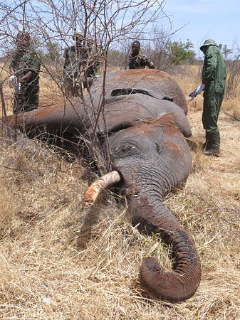
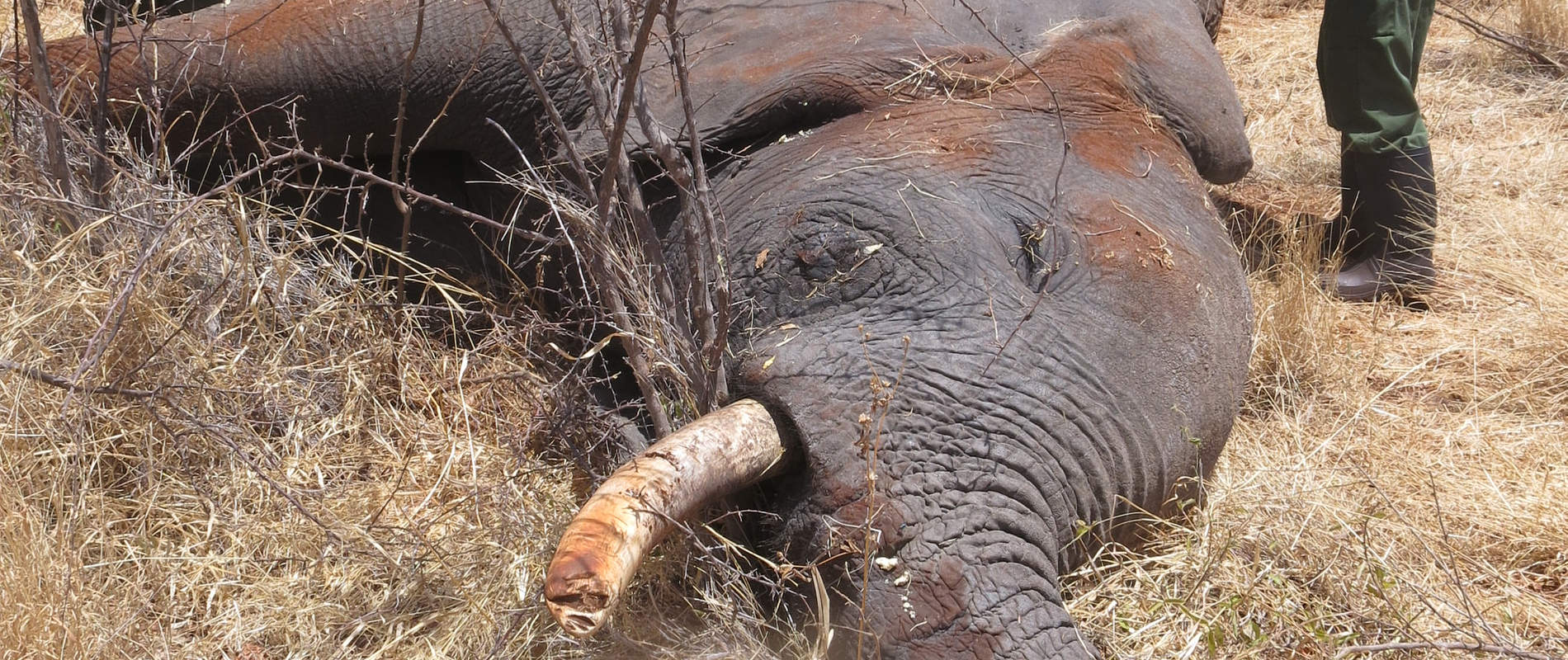
CASE#8: POSTMORTEM OF AN ELEPHANT
Date: 24th February 2015
Species: Elephant
Sex: Male
Age: Adult, 20 years
Location: Bisinadi National Reserve
History
This elephant carcass was reported by KWS rangers on patrol, through their company commander in Meru NP, who requested for a post mortem to determine the cause of death.
An autopsy was done at the scene following standard pathological procedures.
General Examination
This elephant had a rudimentary left tusk; the right tusk was retrieved for safe keeping by the company commander.
Carcass was found on right lateral recumbency with little evidence of struggle at the scene. Carcass inspection revealed old scars on the skin dorsally along the thoracic vertebrae. No other injuries were seen.
On flaying the carcass there were subcutaneous bruises on dorsal and ventral abdomen which may have been self inflicted. There were areas of severe congestion on the muscles of ventral abdomen. In the abdominal cavity, there were parts of the small intestines which were adherent to the omentum and the abdominal wall. Congestion was observed on the peritoneal blood vessels with hemorrhages on visceral surfaces of the gut.


Conclusion
Peritonitis secondary to abdominal trauma
Acknowledgements
We would like to thank the David Sheldrick Wildlife Trust for providing logistical and financial support to enable treatment of these animals.