FIELD VETERINARY REPORT FOR EASTERN CONSERVATION AREA FOR MARCH 2016 Introduction This report describes activities of the Meru veterinary unit in March 2016
FIELD VETERINARY REPORT FOR EASTERN CONSERVATION AREA FOR MARCH 2016
Introduction
This report describes activities of the Meru veterinary unit in March 2016. In Meru National Park two white rhinos were treated for filarial wound while in Solio ranch a Black Rhino was treated for soft tissue injuries. Disease surveillance was also carried out in Mount Kenya wildlife conservancy and a male Hartebeest was relocated to join a group of females in OlPejeta conservancy for breeding.
The unit is supported by the David Sheldrick Wildlife Trust and provides veterinary care to wildlife in northern Kenya.
CASE #1 TREATMENT OF BLACK RHINO ON SOLIO RANCH
Date: 5th March 2016
Species: Black rhino
Sex: Female
Age: 10 years
Location: Solio Ranch
This black rhino was reported to have shown a staggering gait and reluctance to move even when approached on foot or by vehicle. It had also separated from its 2 year old calf. The warden in charge requested for a veterinary evaluation of the rhino to determine the cause of its ill health.


Chemical immobilization and examination
This rhino was darted by vehicle as it was calm on approach. For immobilization we used a combination of M99®5mg with XylazineHcl 60mg in a single 1.5cc DanInject dart syringe with a 2.2 × 60mm needle. The dart was injected into the lateral muscles of the right thigh. Induction time was 8 minutes.


Examination showed lacerations on its external genitalia and hind quarters suggestive of fight wounds. These wounds may have been inflicted during attempted mating and consequent calf separation. Because of soft tissue trauma the rhino was reluctant to move.


Treatment and prognosis
5% Flunixinmeglumine intramuscular
20% Alamycin LA by intramuscular route
Lugols iodine applied on lacerations


This rhino was reported to have recovered following treatment for minor injuries.


CASE#2 TREATING MALE RHINO FOR CUTANEOUS FILARIASIS IN MERU NATIONAL PARK
White Rhino #1
Date: 14th March 2016
Species: White Rhino
Name: Gakuya
Sex: Male
Age: 29 years
Location: Meru NP, Rhino sanctuary
History
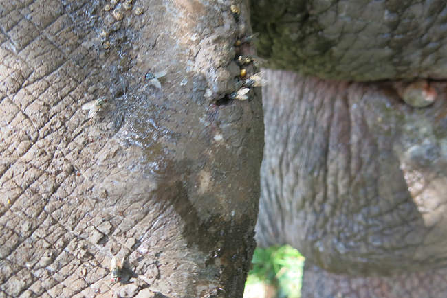
A white rhino was treated in Meru National Park for cutaneous filarial wounds following reports from the rhino monitoring team. Affected rhinos showed extensive wounds on the skin. Treatment was required to prevent wound expansion and infection.
These new cases followed a period of prolonged rainfall in December/ January.
Chemical immobilization and examination
Darting was done from a helicopter. For immobilization we used a combination of M99® 5mg and azaperone 60mg in 1.5cc DanInject dart syringe with a 2.2 × 60mm needle. Darting site was the left gluteal muscle and induction time was 6 and 8 minutes.


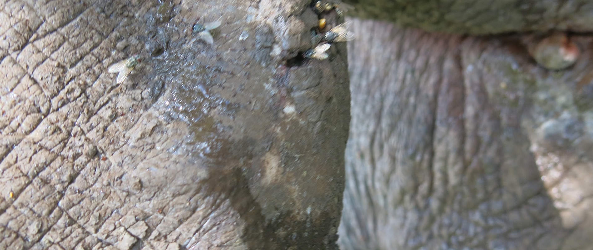
Examination showed ulcerative cutaneous wounds with serrated edges undermined by pockets of pus. Wounds emitted a foul smell due to tissue necrosis caused by bacterial infection. These wounds are characteristic of cutaneous filariasis.


Treatment and prognosis
- Thorough wash with water, dilute hydrogen peroxide and debridement of necrotic tissue. Tincture of Iodine soaked in gauze swabs was the applied
- 1% Ivermectin 300mg administered subcutaneously
- 20% Oxytetracycline administered intramuscularly


Anesthesia effects were reversed within 3 minutes of intravenous injection of Naltrexone Hcl 150mg.


Filarial wounds respond to ivermectin and antibiotic treatment, therefore these rhinos are expected to make a speedy recovery. The rhino monitoring team will observe and report on their progress.
CASE#3 TREATING CUTANEOUS FILARIASIS IN MALE RHINO IN MERU NATIONAL PARK
White Rhino #2
Date: 19th March 2016
Species: White Rhino
Name: Ernest
Sex: Male
Age: 19 years
Location: Meru NP, Rhino sanctuary
History
A white rhino was treated in Meru National Park for cutaneous filarial wounds following reports from the rhino monitoring team. Affected rhinos showed extensive wounds on the skin. Treatment was required to prevent wound expansion and infection.
The Rhino was agitated prior to darting and ran some distance before going down, therefore to manage risk of hyperthermia it was doused with plenty of water. A blind fold was applied to reduce visual stress. It was then roped to right lateral recumbency for examination and treatment.
Chemical immobilization and examination
Darting was done from a helicopter. For immobilization we used a combination of M99® 5mg and azaperone 60mg in 1.5cc DanInject dart syringe with a 2.2 × 60mm needle. Darting site was the left gluteal muscle and induction time was 6 and 8 minutes.


Examination showed ulcerative cutaneous wounds with serrated edges undermined by pockets of pus.


Wounds emitted a foul smell due to tissue necrosis caused by bacterial infection. These wounds are characteristic of cutaneous filariasis.


Treatment and prognosis
- Thorough wash with water, dilute hydrogen peroxide and debridement of necrotic tissue. Tincture of Iodine soaked in gauze swabs was the applied
- 1% Ivermectin 300mg administered subcutaneously
- 20% Oxytetracycline administered intramuscularly


Anesthesia effects were reversed within 3 minutes of intravenous injection of Naltrexone Hcl 150mg.


Filarial wounds respond to ivermectin and antibiotic treatment, therefore these rhinos are expected to make a speedy recovery. The rhino monitoring team will observe and report on their progress.
CASE #4 DISEASE SURVEILLANCE IN MT. KENYA WILDLIFE CONSERVANCY
Date: 16th -24th March 2016
Mount Kenya wildlife conservancy is home to a captive breeding population of endangered mountain Bongo. The conservancy aims to rehabilitate this species for future reintroduction into the wild. A disease risk assessment was carried out in sympatric bush bucks and bovine in the area to inform decisions on species reintroduction. This activity was carried out on 16th to 20th March 2016.
Four bushbucks were captured using the net capture method while three Waterbucks were darted using a combination of M99® and Domitor®. Cattle were restrained using ropes and a crush.


Samples collected include whole blood and serum aliquoted into cryotubes and preserved in liquid nitrogen. Blood smears were also prepared and fixed using methanol and stained for microscopy. Fecal samples were collected from rectum and preserved in ethanol. Ticks on the skin were collected for parasitological studies while hair samples for genetic studies were also collected.


Results from analysis of these samples will provide information on microbial and parasitic disease situation in the area prior to species reintroduction.
CASE #5 RELOCATION OF A HARTEBEEST IN OLPEJETA CONSERVANCY
Date: 27th March 2016
Species: Jackson’s Hartebeest
Sex: Male
Location: OlPejeta conservancy (OPC)
This relocation aimed to introduce a breeding male hartebeest into a group of 14 females in the conservancy and to establish a viable breeding population. These females resided in a fenced off area within OPC which is free from predators.

Capture site was in the main conservancy while the release site was a boma within the conservancy. Immobilization was achieved using a combination of M99®5mg and Stressnil®60mg delivered in a DanInject syringe with a 1.5 × 38mm needle. Induction time was 6 minutes. The hartebeest was blindfolded and loaded onto a truck. The floor of the truck was padded with dry wheat straws to prevent injury and enhance tissue blood perfusion during transport. Transport time was 35 minutes.


Skin lacerations were noted and treated appropriately using povidone iodine. Parenteral 20% Oxytetracycline was given to prevent opportunistic infection caused by transportation stress. This male was successfully released and joined with the females.