EASTERN CONSERVATION AREA VETERINARY UNIT MONTHLY REPORT MAY 2018 Report by: Bernard Rono Summary This report describes veterinary interventions carried out by the DSWT/KWS Mobile Veterinary Unit stationed in Meru National Park in May 2018
EASTERN CONSERVATION AREA VETERINARY UNIT MONTHLY REPORT MAY 2018
Report by: Bernard Rono
Summary
This report describes veterinary interventions carried out by the DSWT/KWS Mobile Veterinary Unit stationed in Meru National Park in May 2018. During this month, rains continued in many parts of northern Kenya hence there was plenty of water and browse for wildlife.
Cases included a hand raised black rhino in Ol Jogi conservancy, a reticulated giraffe in Nasuulu Conservancy, a Grevy Zebra at Buffalo Springs Conservancy and a male elephant was treated for severe lameness in Sabache, Samburu.
We would like to acknowledge support of the DSWT which provides financial and logistical support to the Meru veterinary unit and the KWS management for facilitating the work of this unit in northern Kenya
CASE #1: FIGHT WOUNDS IN A BLACK RHINO
Date: 4th May 2018
Species: Black Rhino
Sex: Male
Location: Oljogi Conservancy
History and clinical signs
This hand raised blind black rhino was attacked by a territorial male black rhino when it was taken out of the boma to forage by its attendants on the evening of 3rd May 2018. The rhino was in pain and discomfort and had not left the boma all day to browse. There were lacerations to the inguinal region with blood loss evident from the blood stained hind limbs, the prepuce was edematous with partial phimosis following blunt trauma. It was immobilized to assess and to treat its injuries.

Immobilization, examination and treatments
The animal was immobilized using Etorphine hcl 3mg and Azaperone tartarate 50mg administered via hand injection in a crush to minimize stress. Butorphanol tartate 5mg was administered intravenously to stabilize the animal once it was recumbent. Vital parameters were monitored closely in 3 minute intervals, the respiratory rate ranged from 4 to 6 breaths per minute and the pulse oxymetry readings for SpO2 ranged from 88% to 94%. The extent of the inguinal injuries was ascertained as superficial lacerations with slight blood loss.


The wounds were subsequently cleaned, debrided and disinfected using water, dilute Hydrogen peroxide and dilute tincture Iodine respectively. Green clay was smeared to hasten the healing process and the wounds were further covered adequately with Oxytetracycline aerosol and fly repellant. The rhino was treated with Amoxicillin trihydrate 15000mg and Flunixin meglumine 1000mg intramuscularly.


Reversal and Prognosis
The rhino was revived using Naltrexone hcl 75mg intravenously at the ear vein. The rhino made a smooth recovery and was up 2 minutes later. This rhino was reported to have shown progressive recovery over the next two weeks. The wounds healed and the rhino was feeding well. The ranch management was advised to keep the rhino within its 100 acre fenced boma to avoid future attacks.
CASE #2: WOUNDS IN A NEWBORN GIRAFFE CALF
Date: 5th May 2018
Species: Reticulated giraffe
Sex: Female
Location: Meru national park
History
Rangers found this giraffe calf and its mother during early morning patrol in the park. It was unable to stand up. An assessment showed deep wounds to the medial aspect of the shoulders which appear to have been pulled apart during parturition by a predator. This calf died a few minutes later from dehydration and shock before any procedures were conducted.

CASE #3: DYSTOCIA IN A RETICULATED GIRAFFE
Date: 7th May 2018
Species: Reticulated giraffe
Sex: Female
Location: Nasuulu community conservancy
History
This giraffe had an obstructed birth, for 48 hours prior to reporting, presented fetal legs and head hanging from the vulva and tenesmus (ineffective straining movements to expel fetus). The vet team immobilized the giraffe to remove the dead fetus and fetal membranes and safe the mother’s life. This intervention was conducted in collaboration with the Veterinary unit in Lewa.

Immobilization, examination and treatment
The giraffe was alone, restless and had lost body condition (body score 1.5 on a scale of 1 – 5) as a consequence of toxaemia from the decomposing fetus. The vet used a lower dosage limit for anesthesia due to its poor body condition. Immobilization was achieved using Etorphine Hcl 8mg and Azaperone tartarate 40mg combined in one intramuscular 3ml Dan-inject dart from a vehicle. Five minutes after darting the giraffe was brought to right lateral recumbence with ropes and physically restrained. Anesthesia was reversed immediately by intravenous injection of Diprenorphine hydrochloride 30 mg through the jugular vein.
On examination we found a dead fetus on anterior presentation with head and front legs hanging from the vulva. The left front leg was flexed at the elbow joint causing obstruction. Fetal parts were partly putrefied.


The vulva was scrubbed liberally with soap and water and lubricated using liquid paraffin. Subsequently the fetus was manipulated to extend the left elbow joint and manually extracted using ropes. Fetal membranes were also removed. Chlortetracycline pessaries were inserted to clear infection intrauterine infection. Amoxicillin trihydrate and flunixin meglumine were administered intramuscularly. The giraffe was then assisted to standing position.


Prognosis
This giraffe is expected to regain its health progressively.
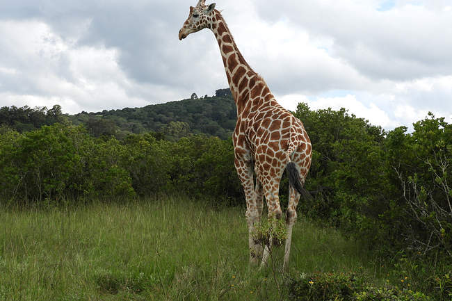
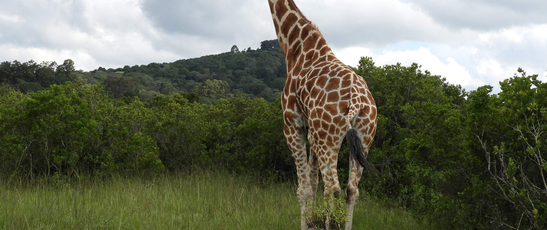
CASE #4: LAMENESS IN A RETICULATED GIRAFFE
Date: 9th May 2018
Species: Reticulated giraffe
Sex: Male
Location: Aberdare Country Club
History
This giraffe in the Aberdare Country Club (ACC) showed progressive lameness for two weeks but the situation had deteriorated prompting need for intervention. The animal presented with weight shifting lameness favoring its right forelimb which had an avulsion wound around the coronary band probably from an injury from rocks in the rocky terrain of ACC.


Immobilization, examination and treatment
The animal was immobilized with Etorphine hcl 10mg and Azaperone tartarate 40mg combined in one intramuscular 3ml Dan-inject dart. The animal was brought down using ropes and restrained physically. Anesthesia was reversed immediately the animal was recumbent using Diprenorphine hydrochloride 30 mg administered intravenously at the jugular.


The wound was approximately 2 inches deep with loss of connective tissue, it was cleaned and debrided to expose the underlying healthy proliferative tissue using dilute hydrogen peroxide and disinfected using dilute tincture iodine. Green clay was smeared to hasten the healing process and the wound covered adequately with oxytetracycline aerosol and fly repellant.The giraffe was treated with 15000mg Amoxicillin Trihydrate and 1000mg Flunixin meglumine administered intramuscularly. It was assisted to standing position using ropes.


Prognosis
A review 7 days after treatment showed reduced lameness and wound healing and the giraffe was on its way to regaining full health.
CASE #5: PREDATOR WOUNDS IN A GREVY ZEBRA
Date: 10th May 2018
Species: Grevy zebra.
Sex: Male
Location: Buffalo Springs
History and clinical signs
The adult male grevy zebra had survived a predator attack but sustained injuries to its right front leg. The male which was alone, grazing intermittently showed discomfort and pain with lameness. There was a large penetrating septic wound on the left lateral aspect of the neck just dorsal to the jugular groove. There was matting of hair in the surrounding neck region.
Immobilization, examination and treatment
The grevy was immobilized using Etorphine hcl 5mg and Azaperone tartarate 80mg combined in one Dan-inject intramuscular dart. The vet darted the zebra from a vehicle.


The penetrating septic wound was around 2 inches in depth and 3 inches in width. It was cleaned, flushed and debrided using dilute hydrogen peroxide soaked swabs to reveal the underneath healthy granulation tissue. The wound was further disinfected using dilute tincture iodine soaked swabs, green clay and opticlox were applied into the dead space before the wound was covered with Oxytetracycline aerosol and wound repellant.The zebra was treated with Amoxicillin trihydrate 7500mg and Dexamethasone 60 mg intramuscularly.


Reversal and Prognosis
The anesthesia was reversed using Naltrexone hcl 100 mg intravenously at the jugular and the animal was up a minute later. Prognosis for recovery is good as the infected wound is expected to heal quickly following treatment.


CASE #6: LAMENESS IN AN ELEPHANT
Date: 27th May 2018
Species: Elephant.
Sex: Male
Location: Sabache, Samburu
History
On 27th May we received a report from rangers at Kalama conservancy of an injured elephant within the community land in Sabache, Samburu. This lone elephant showed lameness of its left hind leg and its mobility was affected due to severe pain. When we visited the area to assess its condition and treat, we arrived there late in the evening due to bad road condition following recent rains.
Immobilization, examination and treatment
Initial assessment showed severe lameness and swelling on the distal part of the left hind leg, no physical injury was seen. Its body condition was fair. It was darted from foot with Etorphine Hcl 12mg in a 1.5 milliliter Dan-Inject dart with the dart placed into the gluteal muscles. Induction time was six minutes after which the elephant was positioned on lateral recumbence for examination and treatment.


Examination showed a large superficial wound (10cm diameter) on the medial aspect of the affected leg with maggots. The cause of the wound could not be determined. The wound was washing with water and dead tissue debrided as much as possible with hydrogen peroxide. Povidone iodine antiseptic and green clay was applied.


Antibiotics and anti-inflammatory drugs were also given via intramuscular route.
Reversal and Prognosis
Twenty five minutes after darting the anesthesia was reversed by intravenous injection of Diprenophine hydrochloride 48mg. The elephant was in standing position three minutes later.
Although there is plenty of water and browse in the area, prognosis for recovery is guarded and it may take a long time to recover. Follow up visit is planned in two weeks if the elephant can be located.