Amboseli Mobile Veterinary Unit (AMVU) attended to six wildlife cases during the month of October 2018. Four animals (an elephant, two zebras, and a buffalo) were treated for different conditions while another elephant and a lion were examined for cause death. The cause of death was human inflicted for both but different in nature for either case, the elephant was killed for ivory while the lion was poisoned in retaliation for livestock invasion.
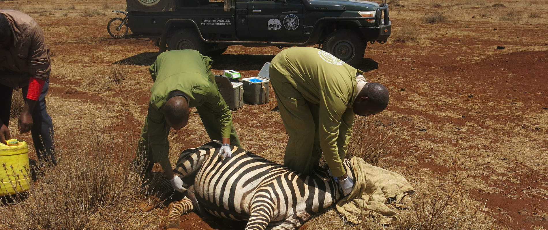
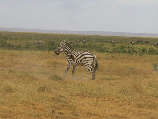
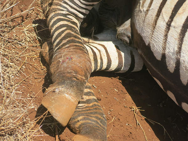
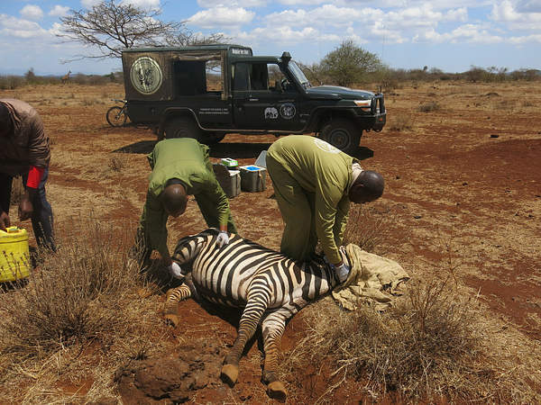
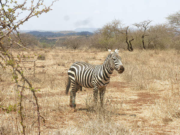
CASE#1 TREATMENT OF A LAME ZEBRA
Date: 15 October 2018.
Species: Zebra (Equus quagga)
Age: Adult
Sex: Male
Location; Amboseli National Park
History
AMVU Team observed a lame zebra while on Park Patrol.
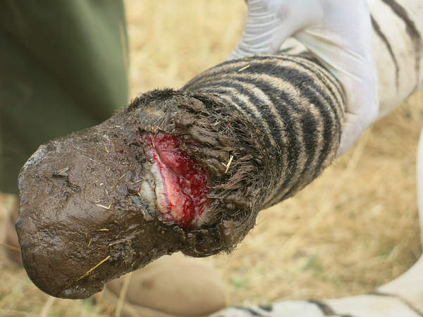
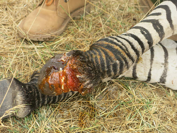
Immobilization, examination and treatment: The zebra was located and darted from a vehicle. A cocktail of 5mg Etorphine Hcl and 70 mg Azaperone immobilized the male to attain complete restraint in 5 minutes after darting.

The right hind leg had a wound slightly above the coronet. The injury affected the skin and part of the common extensor digital tendon attached to the coffin bone. This resulted in lameness and limited mobility. Amoxicillin (Betamox LA®) and Dexamethasone were administered to prevent secondary bacterial infection and pain respectively.
Reversal and Prognosis: Intravenous injection with Diprenorphine revived the animal. Prognosis was good as there were no complications due to the wound. However, due to his lameness the zebra could be easy prey for predators.
CASE#2 TREATMENT OF A WOUNDED ELEPHANT
Date: 20 October 2018.
Species: African elephant (loxodonta africana)
Age: Adult, approximately 50 years
Sex: Male
Location: Tiva River, Tsavo National Park
History: A DSWT pilot while on park air patrol reported the elephant.
Immobilization, examination and treatment: The elephant was located and darted from a chopper. Etorphine Hcl (total dose of 22 mg) immobilized the male elephant. Full restraint was attained in 10 minutes after darting. Treatment was done while the bull rested on lateral recumbency.
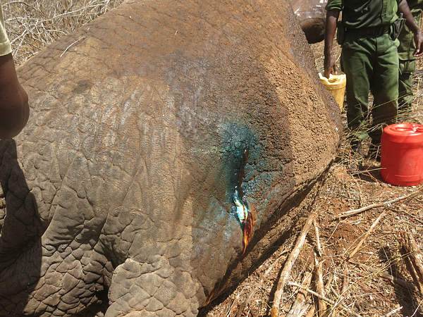
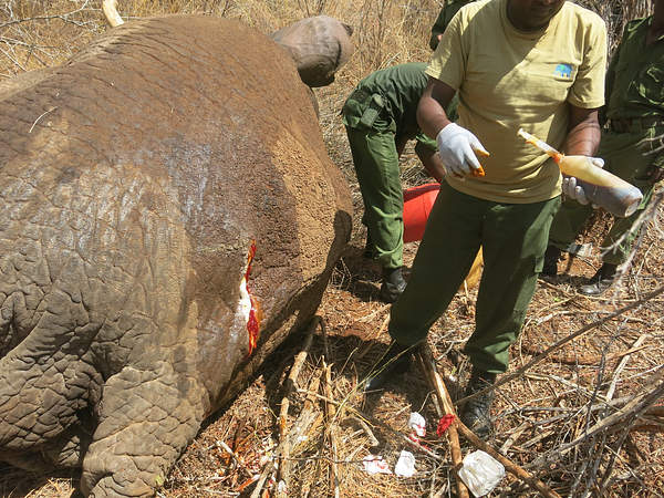
A panga was used to clear the bush around the elephant so that the team could work quickly and easily. A pus filled wound was observed slightly below the backline at hindquarters of the male elephant. Hydrogen Peroxide was used to debride the wound of any old infected tissue. Iodine spray was used to disinfect the wound and green clay was used to cover the wound and prevent any further infection of the wound. Betamox LA®, Amoxicillin was administered to treat for secondary infection.
Reversal and Prognosis: Intravenous injection with 2ml Diprenorphine revived the elephant bull. Prognosis for full recovery is good.
CASE#3 DESNARING A BUFFALO
Date: 22 October 2018.
Species: African buffalo (Syncerus caffer)
Age: Adult
Sex: Male
Location: Salt lake, Taita ranches
History: DSWT rangers reported a buffalo that been captured in a poachers snare on the 22nd of October 2018.
Immobilization, examination and treatment: The buffalo was darted from a vehicle using 5mg Etorphine Hcl and 70 mg Azaperone. Full immobilization of the male buffalo was attained after 10 minutes.
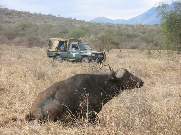
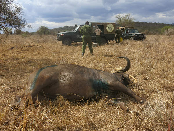
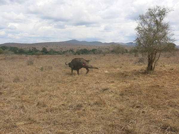
The buffalo was treated with Betamox LA®, Amoxicillin and Dexamethasone. The wire snare was cut with wire cutters, a wound on the chest area was treated with Hydrogen Peroxide and Iodine and the buffalo was released. However, it struggled to stand due to pain.
Prognosis: Prognosis for full recovery is fair.
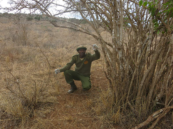
Our mobile vet initiative is in the field every day saving wild lives
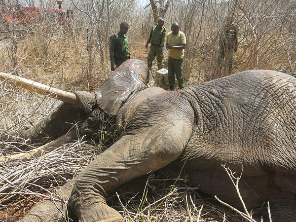
CASE#4 POST-MORTEM EXAMINATION OF AN ELEPHANT CARCASS
Date: 23 October 2018.
Species: African elephant (Loxodonta africana)
Age: Estimated at 35 years
Sex: Male
Location: Kibwezi, adjacent to Athi River
History: George Bakari, Tsavo West, reported a male elephant found dead at Makutano Village in Kibwezi to AMVU on 23rd October 2018. The carcass was examined the same day.
Examination and Findings: The male elephant demonstrated fair body condition. The carcass was fully decomposed. Internal soft tissues were fully destroyed by bacterial activity. Gases and liquids maximally produced and wetted the area around the carcass. The liquid was thoroughly soaked in soil and had dried.
The carcass showed signs of human involvement. Tree branches and leaves covered the carcass in a wooded bush. The carcass had the front of its face cut off, and the tusks were missing and had been visibly removed. Two wounds on the lateral body surface and another on the right fore leg were observed. The actual pre-mortem status of the injury was unclear due to the post-mortem maggot invasion. Trunk tissue had been eaten by maggots and had dried up with neither tissue nor worms. These were all indications that the larvae could have pupated and gone, a process of more than seven days after death. The carcass was deflated, and weight decreased in relation to the volume set by the structural bones.

No flies or eggs on the carcass suggested that the volatile molecules that attract flies were no longer being produced, a natural sign of a more than seven days old carrion. No hyena or vulture activity was observed on the carcass. The carcass was entirely covered with branches and leaves, and that helps explain why the vulture activity was not observed. Lack of either odour or hyena may have led to lack of the same scavenging.
The succession wave of the flies colonizing the carcass indicates an estimated 8-10 days state for the examined carcass. Presence of fully-grown larvae on the body, dried up fluids, the absence of aunts and beetles and a deflated carcass with semi-dried skin would assist in defining the carcases to less than 14 days. Decay odours were reduced and scantly felt. No blow or flesh flies were found on the carcass. However, few houseflies were seen on examiners back. Following the logic of entomological succession on a carcass after death, it is estimated that the carcass was up to 10 days old.
Cause of death: Non-conclusive. The available carcass remain was damaged, decomposed and deficient of a definite cause of death. However, the carcass being covered with branches and missing tusks is evident of a poaching related cause.
CASE#5 POST-MORTEM EXAMINATION OF A LION
Date: 27 October 2018.
Species: Lion (Panthera leo)
Age: Adult
Sex: Male
Location: Olgulului Group Ranch, Loolakirr village, Osewan Area
History: Stephanie of Lion Guardians reported the lion case to the AMVU following a report by guardians Mitiaki Solonka and Lupembe on the 27th of October 2018 at 11:46 am. The two Guardians were investigating vulture activity and three lion tracks when they came upon a carcass of a dead male lion at Osewan area. The guardians identified the dead lion as Lesosio.
Examination and findings: There was little evidence of pre-death struggle; no scratch marks on the ground and no soiling of paws.The male lion carcass demonstrated good body condition before death. It was relatively fresh, and rigor mortis was present but subsiding, an indication that it was within 24-36 hours after death. A small fresh shallow puncture wound over the right humerus was observed but was not related to the death. External parasites, few ticks observed moving over the carcass and were collected. A few flies and small clusters of fly eggs on the ears and body crevasses were observed. However, only a moderate number of flies were present and active.The carcass autolysis process was in process but not sufficiently advanced. The facial area was swollen and stomach bloated. Enzymatic and bacterial activity in the internal organs had started, and gases and liquids produced. However, only little watery, bloody exudate on the ground in front of the nose

A ventral incision opened the carcass, and internal organs exteriorized and observed as follows:
Large areas of ecchymosis (internal bleeding into tissue) were noted in the subcutis and external muscle layers of the neck, chest, and abdomen. Blood vessels with a dark red colour were seen in the skeletal muscles.Trachea: numerous hemorrhages both internally and externally were observed. A frothy bloody fluid found along the trachea, extending to larger bronchi and with small brown particles (possible aspirated vomitus).Oesophagus contained light brown macerated ingesta and had many short hairs. Abdominal cavity macroscopic observations:Peritoneum relatively clean.Spleen enlarged, dark coloured and mottled surface. Stains on the outer surface of the spleen characterize the start of a decomposition process.Liver engorged and intense red colour found with a cooked and mottled appearance.Stomach externalised and opened. A large amount of contents still within the stomach. Contents macerated but with numerous medium sized (5-10cm) pieces of tissue, as well as a lot of short hair.Intestinal lumen filled with air and blackish, greenish liquid paste content.Thoracic cavity: Pericardial sac contents were excessive, watery and bloody. Lungs showed marked congestion. They displayed extensive reddish hemorrhagic areas with a blotchy external appearance.
Cause of death:Respiratory failure, likely due to chocking after forced vomiting. Lack of signs or evidence related to common disease and the macroscopic picture could help explain the likelihood of suspected poisoning.
Recommendation: Laboratory testing: Organ tissue (liver, kidney, intestines, lungs, spleen) and intestinal content collected and stored in fridge at 40 C. Samples to test and quantify for presence of lethal organophosphates and carbamates among other locally available chemical toxicants. Carcass disposed of by burning to avoid environmental contamination. Wood collected and a pyre was built. Carcass dismembered; legs torso, head and placed on top of the fire. Pyre ignited with 10 litre petrol and tended until no discernible body part remained visibleA team from Olgulului Group Ranch (OGR) and lion guardians’ assigned to ensure fire burnt out entirely without the risk of accidental spread.
CASE#6 TREATMENT OF A LAME ZEBRA
Date: 31 October 2018.
Species: Zebra (Equus quagga)
Age: Adult
Sex: Male
Location: Isinet, Next to Kimana Sanctuary,
History: A report of a lame common zebra was received at AMVU on 31 October 2018 from Big Life Foundation Rangers. The zebra was grazing at Isinet area near Kimana Sanctuary.
Immobilization, examination and treatment: The zebra was located and darted from a vehicle. Etorphine Hcl (total dose 5mg) and 60mg Azaperone immobilized the male zebra. Successful restraint and lateral recumbency was attained in 6 minutes after darting.

The Zebra suffered an old injury that led to the fracture of the femur bone on the right hind leg. The zebra used three legs and could barely step down with the lame leg. The leg had healed but the fractures bones had joined together nicely. 20ml Betamox LA®, Amoxicillin was administered to prevent secondary bacterial infection. Flunixine was used for pain relief. Iodine was sprayed into the open wound.
Reversal and Prognosis: Diprenorphine reversed the animal from the effects of Etorphine. Prognosis for full recovery is fair since the injury has complicated the zebra’s use of the limb. It is likely that the equine would easily fall prey to a predator.
OTHER OBSERVATIONS
Many elephants have left the Park for Community Ranches. Only few greater flamingoes were present in the Amboseli waters as opposed to earlier sightings in August and September 2018.
Acknowledgment
Report by KWS Vet Dr Kariuki. We thank David Sheldrick Wildlife Trust (DSWT) for material support and Mobile Unit funding, KWS for logistical support and security. DSWT and Big Life Foundation rangers for reporting and monitoring of distressed wild animals. We are always grateful for your help in conservation in Kenya.