Introduction The veterinary activities in the Central Rift region and Masai Mara went on successfully during the month of November, 2008
Introduction
The veterinary activities in the Central Rift region and Masai Mara went on successfully during the month of November, 2008. Some of the activities that took place include capture and sampling of wildebeests and gazelles for (mange) skin disease. African swine fever disease surveillance in Ruma National park also continued and two more bush pigs were captured and sampled. Collaring of one adult male elephant in Ngurumani area in Magadi to assist scientists and wildlife managers in monitoring the movement of elephants and control cases of human-wildlife conflicts in the area. Clinical cases that were attended to in November include treatment of common zebras which were either injured or entangled by snares in Naivasha area. Rescue and treatment of a giraffe which had an arrow head on the back in Keekorok area in Masai Mara. The veterinary unit still lacks a refrigerator for storing biological samples, lack of internet access and inadequate power supply are some of the challenges facing the vet unit in Mara.
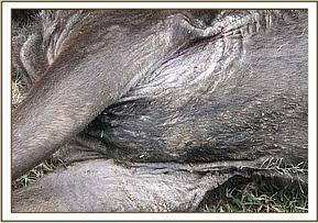
Treatment and samples collection from a wildebeest with mange infestation in Mara Triangle.
Some of the wildebeests calves in Mara were reported to have mange infestation along Mara river near the Mara Bridge. The disease was suspected to have contributed to deaths of some calves and could be easily transmitted to cheetahs which feed on those wildebeest calves. The veterinary unit in Mara intervened and managed to get at least one wildebeest calf with heavy infestation on the neck and head region.
The animal was captured by chemical immobilization through darting using 3mgs of etorphine hydrochloride combined with 20mgs of Xylazine hydrochloride and it took 5 minutes for the drug to take effect. Skin scrapping samples from the skin lesions were collected and stored in 70% ethanol. The animal was treated with Ivermectin administered through the sub-cutaneous tissue. Other treatments using antibiotics and tincture of iodine applied on the infected skin. The anaesthesia was then reversed using 12mgs of Diprenorphine hydrochloride combined with 5mgs of Atipamezole hydrochloride. 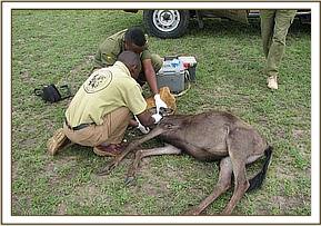

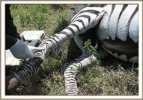
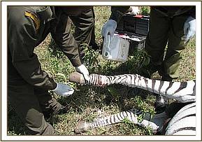
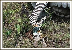
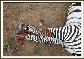
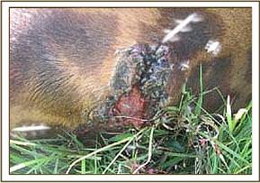
Removal of a snare and treatment of a common zebra in Kigio Wildlife Conservancy in Naivasha.
This was a case of a male adult zebra that had been sighted with a deep and severe wound circumventing the fetlock region of the right hind-limb, it had stayed with the injury for a few days and was unable to move and graze at the same rate with other zebras. Through the assistance of the conservancy rangers, the vet team was able to locate the animal in a fairly open ground. It was then captured by chemical immobilization through darting.
The wire was then retrieved from the wound and removed completely leaving an extensive raw wound on the limb. The wound was also treated using 10% hydrogen peroxide then a tincture of iodine applied topically on it. Anaesthesia was then revived and the animal released back to the wild after being treated successfully. 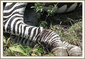

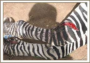
Treatment of an injured common zebra in Game Sanctuary in Naivasha
The zebra was located in Game sanctuary with a deep cut on the right front limb, the wound was still fresh with blood oozing out, the cause of the injury was not ascertained. The zebra was the captured normally by use of chemical immobilization and treated using antibiotics, antinflammatory drugs and topical application of tincture of iodine. It was then revived from anaesthesia after the treatment. It had better chances of healing since the wound was still fresh and treated at the right time before it developed infection. 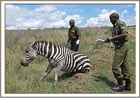

Treatment of a giraffe with an arrow head on the back
This was an adult male giraffe that was sighted near Keekorok lodge by County council rangers and reported to the veterinary unit in Mara, it had an arrow head sticking on the back. It had stayed with this for sometime and the wound created had developed some infection.
The giraffe was darted using 13mgs of etorphine hydrochloride combined with 30mgs of Xylazine hydrochloride and took about 6minutes for the drug to take effect. The arrow head was then removed from the back region near the lumbar-sacral joint. The wound was also cleaned and treated with hydrogen peroxide and a tincture of iodine applied topically, more antibiotic drugs were administered intramuscularly. 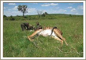
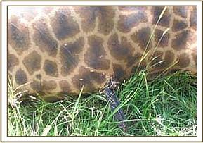
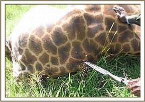
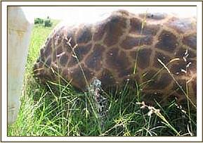

The animal was then revived from anaesthesia and released feeling relieved. Prognosis was good because the foreign material that was causing much pain and irritation had been removed and the wound was showing some signs of healing already. It was not known where the giraffe was speared from because it belonged to a resident herd that do not migrate outside the reserve, could be there were some people hunting for game meat and happened to get into the reserve and speared the animal. The management of Mara game reserve were informed duly and will find out.
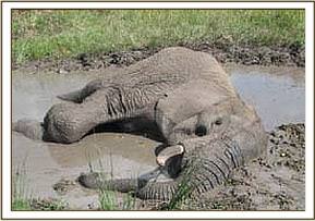
An elephant with a fractured leg in Mara near Makari bridge
The elephant was found lying in a small pool of water near Makari bridge, it was unable to stand up or walk. It was a sub-adult female elephant of about 10 years old, the rest of the family had left her behind since it was completely unable to move. The veterinary team while being assisted by the Mara rangers and staff from Keekorok lodge managed to pull the elephant out of the pool using ropes and a vehicle. It was later found to have a fractured right front limb, a complete but closed fracture of the humeral bone. The animal was weak and harmless so we could manipulate it safely without any form of anaesthesia.
The fracture could not be corrected because of the heavy weight of the elephant, it was under intense pain and had very little chance of survival, it was also becoming an easy target for poachers while it remained lying motionless in the pool, so the decision was made to euthanize it humanely and recover the tusks. The carcass was then pulled and taken far away from the water place, the tusks were removed and kept safely by the management of Mara game reserve awaiting submission to KWS stores. 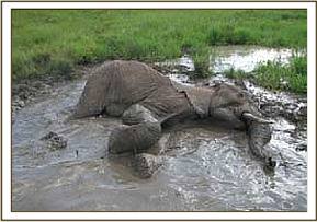

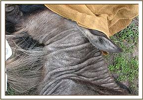
Investigations for cases of skin disease (mange) in cheetahs, Thompson’s gazelles and wildebeests in Mara.
Mange is a highly contagious skin disease caused by mites (Muller. et. al., 1989).Various genera of mites have been documented but the commonly found mites in cases of mange are Sarcoptes and Notoedric mites. Mites affect domestic animals, humans and wildlife (Pence and Uckermann, 2002). Mange is also a disease of zoonotic importance.
The cheetah (Acinonyx jubatus) population in Kenya is estimated to be 793 individuals (Gros, 1998). The species is listed as endangered and is in Appendix 1 by the Convention on Trade in Endangered Species of fauna and flora (CITES). Diseases are among the major factors that have led to the decline in population and mange being placed among the leading causes of death (Weber & Rabinowitz, 1996).
The disease may reach epizootic proportions in certain wildlife populations such as cheetahs in Africa. It was first reported in Cheetahs of Masai Mara by Mwanzia et. al., 1995). It is believed that Cheetahs do get infected through contact with Thompson’s gazelles or any other herbivores which is their main prey in the wild.
The net effect of a mange epizootic can have serious consequences in remnant or fragmented populations of CITES – listed, threatened or endangered species where loss of individuals through death can be critical to the survival or restoration of a species. There have been continuous investigations, monitoring and treatment of mange in cheetahs of Masai Mara since the year 2000 in an attempt to control the disease.
The Veterinary staff in Masai Mara has since been following all the cheetah and other wildlife population to treat any animal with skin infections and collect skin scrapping samples for laboratory analysis and investigations on the epidemiology of the disease in Mara. During the month of November, 2008, one male wildebeest was found to have skin lesions suspected to be caused by mange. The animal was captured by chemical immobilization, skin scrapping samples collected and treated for mange and released. 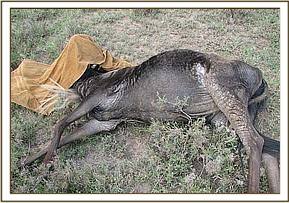

Several gazelles and livestock in the immediate surroundings of Mara reserve were observed and data on disease prevalence and distribution collected for analysis.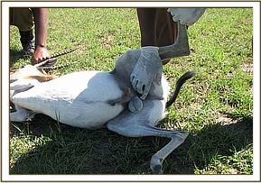
AFRICAN SWINE FEVER DISEASE SURVEILLANCE IN RUMA NATIONAL PARK:
Introduction
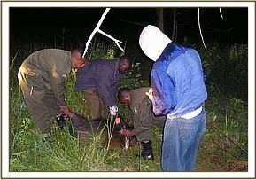
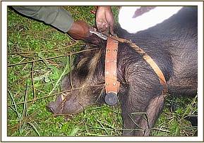
Kenya Wildlife Service (KWS) veterinary department in collaboration with International Livestock Research Institute (ILRI) initiated African swine fever (ASF) disease surveillance in Ruma National park since November, 2007. The surveillance work has been conducted in the park three times since its initiation last year. During the month of November, 2008, two bush pigs were captured at night using capture nets one was fitted with a GPS collar and blood and tissue samples collected from both the pigs and released. The operation lasted for 12 days and it was successfully conducted. 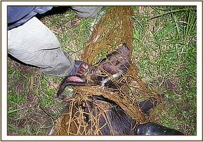

Background information about ASF
African swine fever (ASF) is a highly contagious disease of pigs, first described in Kenya by Montgomery (1921). It is caused by a large DNA virus and is considered to be one of the most complex viral diseases to affect domestic animals. The disease is endemic in many African countries south of the Sahara desert. In Europe it is still endemic in Sardinia (Italy) and outbreaks were confirmed in the Alentejo region of Portugal in November 1999 (OIE, 1999). The disease has been successfully eradicated throughout the rest of Europe. ASF is classified in list A of the Office International des Epizooties (OIE, 1999) and inflicts significant socio-economic impact on affected countries.
Reservoir hosts and transmission of (ASF)
The ASF virus affects both wild and domesticated pigs and is transmitted by soft ticks of family Argasidae, genus Ornithodoros. In Africa, it is mainly transmitted through Ornithodoros moumbata in Africa (Plowright et al., 1970) and Ornithodoros erraticus in the Iberian Peninsula (Sanchez Botija, 1963).
Ornithodoros corinaceus, a tick indigenous to the USA, has also been found to harbour and transmit ASF virus in experimental settings (Groocock et al., 1980), as has Ornithodoros savignyi which is present in Africa (Mellor and Wilkinson, 1985).
In Africa, ASF virus usually induces a non-apparent infection in three wild boar species: warthog (Phacochoerus aethiopicus), giant forest hog (Hylochoerus meinertzhageni) and bush pig (Potamochoerus porcus). Infection is characterized by low levels of virus in the tissues and low or undetectable levels of viremia. In the case of bush pigs P. porcus, viremia has been observed between 35 and 91 days following infection and the virus can persist in lymphatic tissues for 34 weeks (Anderson et al., 1998). Viral infection normally moves from these animals to domestic pigs through a biological vector, Ornithodoros moumbata, and not by direct transmission.
ASF virus infections in the African vector Ornithodoros moumbata are transmitted by transovarial and transtadial routes, whilst only transtadial transmission has been observed in the European vector Ornithodoros erraticus.
Significance and objectives of ASF surveillance in Ruma National park.
African Swine fever (ASF) is classified on OIE list A, which by definition contains only diseases that have the potential for very serious and rapid spread, producing serious socio-economic consequences (OIE, 1999). The last five years (1985-1990) of the ASF Spanish eradication programme cost approximately US $92 million.
Communities living around Ruma National park do keep pigs which are slaughtered normally for food especially during festivities. African swine fever is a disease of pigs but can induce a non-apparent infection in wild pigs such as bush pigs which then act as reservoir hosts for the disease and may transmit the virus to the domestic pigs.
The main objectives of ASF surveillance is to confirm if the bush pig population within the park have ever suffered from the disease by conducting serological tests to detect ASF antibodies in serum.
To test some of the diagnostic kits which have been developed for diagnosis of ASF in both domestic and wild pigs.
If the virus is found to be circulating in Ruma bush pigs then control measures can be put in place to avoid transmission of the disease to domestic pigs that can lead to an outbreak and serious economic losses due to pig mortalities in Kenya.
Methodology for bush pig capture and samples collection
Bush pigs are nocturnal animals that can only be seen late in the night, they are also extremely shy and cannot come close to humans. During day time they hide in deep thickets where they cannot be seen. These factors make it difficult to capture and handle bush pigs for sero-surveillance. In Ruma National park, they are commonly sighted near garbage collection sites within the residential areas at the park headquarters, forest department residence, .Nyatoto gate, and Wiga gate. They are always seen at night while invading crop farms at night away from the park then hide in the nearby forest during day time. The use of capture nets is the most reliable method applied in Ruma.
Samples collection
Blood samples were collected using vacutainer needles and tubes from the femoral vein and coccygeal vein then drawn into 14ml plain tubes coated with clot retractor and EDTA coated tubes and put in a cool box overnight. Ticks were picked from the skin of the animal using rat-toothed thumb forceps and placed in small containers and placed in a cool place. Tissue samples were also collected by cutting a small portion of the ear-pinnae.
Samples storage and processing
The blood samples were centrifuged at 1500 rounds per minute for 15 minutes then serum extracted into cryovials and kept in a deep freezer for submission to the lab for analysis. Tick samples were also kept a deep freezer, tissue samples were dissolved and kept in a freezer.
Prevention and Control of (ASF)
Control of the disease is usually achieved by the control of tick vectors. No treatment or vaccine is available for ASF and control of this disease is based on rapid laboratory diagnosis and the enforcement of strict sanitary measures.
Depending on the epidemiological status of the disease in a particular region, different measures are recommended. In endemic areas of Africa, the most important factor is to control the natural tick vectors and wild pig reservoirs, and/or limit their contact with domestic pigs. The identification and slaughter of sick and carrier animals is crucial to the control of the disease.
Conclusion
During the month of November, the veterinary unit achieved much in terms of handling clinical cases as well as disease surveillance and investigations in Central Rift region. Basic laboratory equipment were provided by the David Sheldrick Wildlife Trust but the unit still requires a laboratory technician based in Mara to help improve the veterinary services and research activities in the region.
Reported by: Dr. Domnic Mijele