The month was characterised by windy dry days with insignificant precipitation. Fodder for browsers is still available with most of the migratory wildebeests having gone back to the Serengeti. Fewer cases were reported and handled during the period as compared to the previous month. Three rhinos and an adult elephant cow were lost within the Mara Triangle Conservancy, all in close proximity and within a short span. Samples were however collected and are under analysis.
CASE#1 INJURED LION
Date: 12th November 2018
Species: African lion
Sex: Male
Age: Adult
Location: Olkinyei Conservancy
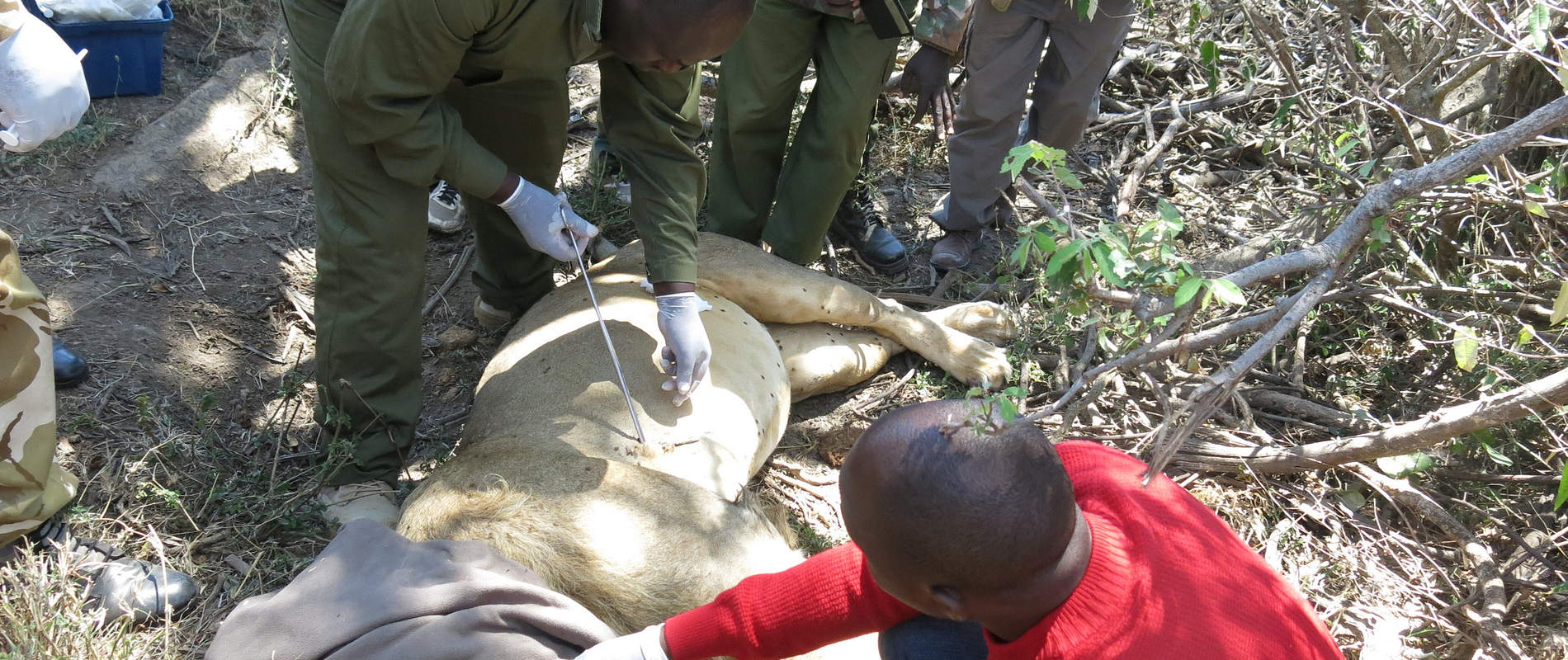
History: This lion was seen with an arrowhead sticking out from his right shoulder by Porini Cheetah Camp managers with their guests, whilst on a game drive. They called the Veterinary Unit for assistance and informed the Conservancy Patrol teams to monitor. This lion was found lying in the shade of a small bush having recently fed. The arrowhead could be seen sticking out from his right shoulder.

Immobilization, examination and treatment: Restraint was achieved chemically by use of a combination of 6mgs Medetomidine Hydrochloride and 250mgs Ketamine Hydrochloride delivered through a 3ml daninject dart. Darting was done from a vehicle with the drugs taking full effect after ten minutes. Examination revealed an arrowhead lodged on his right shoulder with sepsis setting in. It was estimated to have been in place for more than three days.
Fortunately this was not a poisoned arrow. It was gently removed with the resultant wound being debrided with the help of Hydrogen Peroxide and gauze swab. Rinsing was done with copious amounts of water before the wound was disinfected with Tincture of Iodine. Oxytetracycline wound spray was then applied topically. Other treatments given include the administration of 3000mgs Amoxicillin antibiotic and 16mgs Dexamethasone Sodium anti-inflammatories.
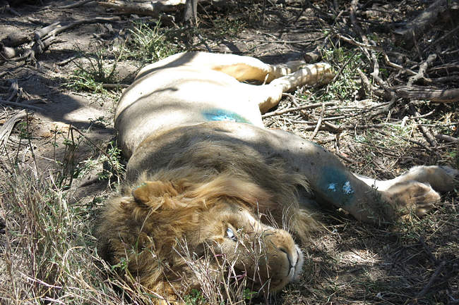

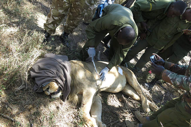
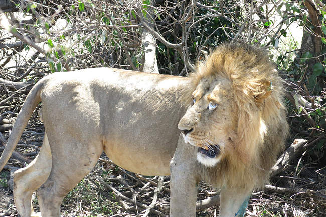
Reversal and prognosis: Reversal was achieved by use of 15mgs Atepamezole Hydrochloride given intramuscularly one hour after immobilization. He woke up from anaesthesia and retreated to a small thicket. Prognosis for full recovery is good.
CASE#2 EXAMINATION OF A BLACK RHINO CARCASS
Date: 12th November 2018
Species: Black rhino (Diceros bicornis)
Rhino ID: 1552 (Sairowua)
Sex: Male
Age: 37 years
Location: Mara Triangle Conservancy
History: The carcass of this male black rhino was discovered the morning of the 12th of November, by Mara Triangle Conservancy rangers on patrol. They positively identified the carcass as belonging to one of rhinos known to live within their Conservancy christened ‘Sairowua’. They informed the Mobile Veterinary Unit and requested a post mortem to establish his cause of death. History from the rhino warden indicated his body condition was satisfactory with a score of 3 out of 5 (in a scale pf 1-5 where 1 is poor and 5 good). He was reported to be among the oldest rhinos in the Conservancy.
General observation and important findings of the carcass: This was an extensively scavenged and partly dismembered carcass.Carcass estimated to be 5 days old. He was found lying on his left lateral position. There were no signs of struggle before death at the scene. All the soft tissues and internal organs were missing, thought to have been consumed by scavengers, mainly hyenas and vultures. This left a shell of skin and bony tissues. Infesting maggots could still be found.
Apart from scavenged areas, no fresh injury could be detected on any part of the skin. The ears and both the upper and lower lips had been ripped off. The skull was intact with no injury detected. The right lateral surface and ventral abdomen had old and healed scars thought to have been caused by cutaneous filariasis. The molars and pre-molars showed advanced degradation associated with age.
Both the anterior and posterior horns were found apart and a few meters from the carcass. The anterior horn on stable equilibrium position and the rear smaller horn on neutral equilibrium. One hoof was found close to the bigger horn and all of them bore bite and scratch marks suspected to be by hyenas. Examination of the attachment position of the horns on the carcass’s rostrum showed the horns became loose and possibly fell off during the decomposition process or easily pulled out by scavengers. There were no signs of forced extraction or excision. No evidence of bleeding around the attachment area or the ground beneath.
On opening the carcass, nothing much could be captured grossly due to its state and interference from the scavengers. Skin tissue was collected for DNA analysis and banking.

Conclusion: At 37 years and with the history gathered from the Rhino Warden on his condition, this rhino was not doing very well given that there is plenty of browsing material at the moment in the Conservancy. Owing to the state of the carcass (advanced decomposition and scavenging), the post-mortem could not conclusively reveal the actual cause of death but geriatric complications could have contributed to his death. Both horns were recovered dispersed within the vicinity of the carcass, examination revealing that their displacement from the carcass was an act of the scavengers, particularly hyenas. The findings highly suggest that this rhino could have died of natural causes.
CASE#3 EXAMINATION OF AN ELEPHANT
Date: 15th November 2018
Species: African elephant
Sex: Male
Age: 20 years
Location: Oloisukut conservancy
History: This young bull was seen alone and moving with difficulty by Oloisukut Conservancy Rangers. They called the Mobile Veterinary Unit for assessment.

General observation and diagnosis: This elephant was found isolated from the rest of the herd standing near a watering point. He appeared to have difficulties swallowing as he regurgitated much of the water he tried to drink. The trunk was extended and kinked to the right side and so was the tail. He also appeared to drag his left fore and hind leg on movement. These signs were consistent with left ipsilateral paralysis, a sign that the right side of his brain had problems.
The team felt immobilization would further complicate the situation of this elephant and was withheld. Damages to the brain heal slowly and given that this elephant could still walk, close monitoring was advised to see how he will cope with the challenge before next action is taken. It is reported he is still moving around and feeding intermittently though with some difficulty. He is being closely monitored by rangers. He can regain a great percentage of his health with time.
Our mobile vet initiative is in the field every day saving wild lives
CASE#4 POST-MORTEM OF A BLACK RHINO
Date: 16th November 2018
Species: Black rhino (Diceros bicornis)
Rhino ID: 1562 (Naiteru)
Sex: Female
Age: 16 years
Location: Mara Triangle Conservancy
History: The carcass of this young female was seen the morning of the 16th of November by Mara Triangle Rhino Patrol Team. They positively identified her as Naiteru (ID 1562). She is reported to have had two calves with the youngest about two and a half years old still accompanying her. They sought the services of the Mobile Veterinary Unit to establish the cause of her death. This was the second rhino carcass to be found within the same general area in a span of four days. The previous carcass of an old male black rhino had been extensively scavenged on leaving a shell estimated to have been 5 days old. The latest was however a relatively fresh carcass estimated at two days with partial scavenging.
The distance between the previous and the current carcass is about 4 kilometres with reports that their territories were overlapping. There were reports that an old elephant carcass was recovered a fortnight ago near a salt lick several kilometres away but within the same Conservancy. The tusks were still intact. The frequency and proximity of the two carcasses has created a lot of concern with the Teams currently carrying out intensive patrols to confirm status of the other rhinos. Information gathered reveals that other herbivores (grazers and browsers are thriving well in the conservancy).
General observations and significant findings: This rhino was found on right lateral recumbency with early putrefaction. No external injury was detected even with the carcass being turned over.
Other general findings were:
There were signs of struggle before death at the scene.Severe emphysema with early maggot infestationBoth horns were intact.This rhino appeared to have been in good body condition before death. Estimated scale of 4.5 out of 5 in a scale of 1 -5 ( 5 being perfect and 1 being poor)Carcass was estimated to be two days old.Part of perineum and posterior ventral abdomen had been ripped off by scavengers accessing the lower gastrointestinal tract whose contents had spilled to soil the peritoneum.Her left ear, upper and lower lips together with apex of the tongue had been mauled.
On opening the carcass, the following findings were noted:
The spleen and both kidneys were missing, scavengers having pulled them off and consumed them.There was extensive subcutaneous haemorrhaging, more pronounced on her ventral chest area. The lower gastrointestinal tract had been punctured with contents floating freely within her abdominal cavity. The liver had a cooked appearance with advanced autolysis noted.The diaphragm bore perforations likely caused by scavengers. The lungs were congested and frothy.
The pericardium was intact with the heart appearing normal in size and gross architecture. The oesophageal mucosa was peeling off, this being expected post mortem change. Trachea bore some secretions extending from bronchi and bronchioles and further down the respiratory tree. This rhino was gravid with an eight old male calf which was fresh and could have died soon after the mother’s death. The skull and its contents appeared normal. All other organs appeared normal with expected post mortem changes noted in them.

Conclusion: This rhino appeared to have died struggling. The frothy lungs and tracheal secretions show that there was lung congestion and that she had difficulty breathing. Due to advanced putrefaction, no viable bacteriological samples could be collected. However, liver tissue samples and stomach contents were collected and will be presented for toxicological analysis. Given that this rhino shared the same niche with the recently lost male, environmental samples from areas of common use (salt lick) were collected and will be presented together with the other samples for analysis and comparison.
CASE#5 RHINO POST MORTEM
Date: 19th November
Species: Black rhino (Diceros bicornis)
Rhino ID: (Clean) Kantai
Age: 3 years
Sex: Male
Location: Mara Triangle Conservancy
History: Following the discovery of two rhino carcasses in a span of 3 days in Mara Triangle Conservancy, there was concern and urgent need to account for all the rhinos inhabiting the conservation area. The Conservancy deployed personnel with aerial support to do a complete recce in order to locate and confirm the status of each of the rhinos. It was during the exercise, when the carcass of this young male christened ‘Kantai’ was discovered in the afternoon of 19th November 2018.
He did not have any identification number given that he had not been ear notched. He was positively identified as the youngest calf to the female that died a few days prior. It is reported he stayed close to his late mother and shared a common niche. This rhino was reportedly seen healthy and in good shape two days prior to the discovery of the carcass. An autopsy was required to shed light on possible causes of his death and a decision was to be made on the way forward. A team from Headquarters Veterinary Department joined hands with the resident Veterinarian to carry out an autopsy and collect relevant samples.
General observation of the carcass: The carcass of this rhino was found on left lateral recumbency moderately bloated. The carcass was estimated to be 36 hours old. Lions were found scavenging on the carcass with both upper and lower lips together with the perineum and the tail mauled. Early stage maggots were beginning to infest. There was evidence of struggle before death at the scene. He appeared to have been in perfect body condition before death.
Subcutaneous emphysema was noted. Apart from those wounds occasioned by scavenging, no other external injuries were seen.Rigor was present though gradually dissolving. Use of metal detector to scan the entire carcass was negative for metallic foreign objects. There were fully formed rhino faecal boli with good consistency at the scene of death. It appeared the faeces were released at the point of death. Both horns were present.
On opening the carcass, the following findings were noted: Extensive subcutaneous haemorrhages were seen on the ventral side of his chest. Other prominent haemorrhages were seen along the spine.The lungs had undergone advanced autolysis. The heart had some zones that appeared autolyzed. The liver and the spleen were completely autolyzed. Kidneys had cooked appearance but had maintained their normal shape. The entire gastrointestinal tract was full with evidence this rhino had not stopped feeding and his death was acute. The stomach was full with the mucosa easily pealing out.The smaller intestines appeared haemorrhagic with evidence of erosion. The lower gastrointestinal tract had ingesta, with the lumen easily breaking on palpation. The mucosa had dark zones.

Conclusion: This rhino occupied the same niche with the previously lost rhinos. They appear to have died under similar circumstances, with their deaths being acute. Differentials of acute deaths in rhinos which include clostridial infections and possible exposure to toxic or poisonous substances were considered. Stomach contents, liver and kidney tissues were collected for toxicological analysis.
Intestinal tissue was also collected for histopathology and clostridial toxin examination.
CASE#6 SICK CHEETAH
Date: 20th November 2018
Species: Cheetah (Acinonyx jubatus)
Age: Adult
Sex: Female
Location: Mara Triangle Conservancy
History: This cheetah was being monitored by Mara Triangle rangers for two days and she appeared to be losing condition. They called the Mobile Veterinary Unit for assessment and intervention.

General observation and way forward: She was found lying under a small shade severely emaciated but with good demeanor. Immobilization was ruled out due to her condition as the Team felt she might not withstand anaesthesia. She appeared to have recently developed diarrhoea that had since stopped. She was given 450mgs Clindamycin antibiotic intramuscularly through remote delivery with a 3ml daninject dart.

In addition she received a muscle tonic at the same time to improve nerve and muscle co-ordination. This darting was done from a vehicle. She moved a short distance before dropping the dart, which was collected.
Prognosis: Prognosis is guarded. The rangers were advised to monitor her and report the progress.
CASE#7 INJURED ELEPHANT
Date: 22nd November 2018
Species: African elephant
Sex: Female
Age: Adult
Location: Siana Conservancy
History: This elephant was seen by Elephant Aware rangers at the bottom of a cliff seemingly injured. They reported this to the Mobile Veterinary Unit.
General observation and way forward: This elephant was found on left lateral recumbency struggling to pull herself up, with the trunk and forelimbs active. It appeared she had been in this position for at least three days. She was in a shallow pool having fallen from a cliff estimated at ten metres. It appeared there was an attempt on her life through use of poisoned arrows and she ended up falling down the cliff as she escaped. She seemed to have damaged her spine with resultant posterior paralysis as she could not move her hind legs and tail.

Two lodged poisoned arrows were seen on her withers area and off the cervical part of her spine. Another arrow injury was seen on her lower back. An attempt to pull her up using ropes and vehicles failed to materialise as her hind legs were completely paralysed. After several hours of attempt, it was clear this elephant could not make it owing to spinal damage.
This presented a poor prognosis and the team felt the need to stop her further suffering. Euthanasia was advised and was effected immediately. The tusks were retrieved and taken for safe custody by KWS security personnel.
CASE#8 POST-MORTEM OF A DEAD ELEPHANT
Date: 25th November 2018
Species: African elephant
Sex: Female
Age: Adult (Approximately 40 years)
Location: Mara Triangle Conservancy
History:
This elephant was seen dead the morning of the 25th of November by Mara Triangle Patrol Rangers. Three rhino carcasses were found in the same area over the last two weeks whose cause of death had not been established so far. The Mobile Veterinary Unit responded to carry out a post mortem examination and collect samples for analysis.
General observation of the carcass: The partly scavenged carcass was found on right lateral recumbency with abdominal organs partly exteriorised by scavengers. Perinium and the trunk had also been ripped off by predators, among them lions who were seen close by. No other external injuries were noted with negative result on scanning for foreign metallic objects. She appeared to have died suddenly. Further assessment revealed an elephant that was in satisfactory body condition and at end stage nursing. The carcass was estimated at less than 24 hours old with rigor slowly dissolving and with signs of struggle at the scene before death. There was notable swelling of the head all the way to the neck. Blood appeared dark but clotting.

On opening the carcass, the following findings were noted: The lungs appeared slightly congested with clear trachea and bronchi. The heart was normal in shape, size and texture. The right ventricular endocardium was haemorrhagic. The entire epicardium had petechiae. Oral mucosa and the tongue appeared cyanotic. Spleen was slightly enlarged. The liver was severely congested and friable.Stomach was full with its mucosa easily peeling off. The small and large intestines appeared slightly eroded but full with ingesta. Rectum had fully formed faeces with easily peeling off mucosa. The kidney appeared normal in size and consistency.
Samples from the liver and kidneys were collected for histopathology and toxicology. Stomach contents were collected for toxicology while intestinal tissue was collected for microbiology, toxicology and histopathology. Impression smears were prepared for microscopy.
Conclusion: This appeared to be a sudden death with the differentials of acute death being considered. This elephant died at the same general area where three black rhinos died in a period of less than ten days. This shows there could be a common source that could either be a toxin in the environment of access or a virulent pathogen shared by the species. Samples collected might give an indication on the problem currently being experienced in the conservation area.
Case#9 Examination of an Elephant Carcass
Date: 27th November 2018
Species: African elephant
Age: Adult
Sex: Female
Location: Naboisho conservancy
History: The carcass of this elephant was seen by tour guides at Naboisho Conservancy who informed the Conservancy Management. The Mobile Veterinary Unit was then called to examine this carcass and possibly determine the cause of death.
General observation and findings on the carcass: This elephant was found on left lateral recumbency, both tusks intact and were retrieved for accounting and safe custody by the Conservancy security personnel. The age of the carcass was estimated to be 3-4 days.Scanning with a metal detector was negative for metallic foreign object She appeared to have been in good body condition before deathThe scene of death had been tampered with extensively by scavengers.Many hyenas and vultures were found feasting on the carcass with only skin and bony tissues remaining.
Conclusion: This carcass had been extensively scavenged on with only a shell of skin and bony tissues remaining. All soft tissues had been consumed by the scavengers. Abdominal contents were missing as they had been dispersed far and wide by the scavengers as they scrambled for the carcass. As such no meaningful samples could be collected and the cause of death could not be grossly determined.
Acknowledgement
Report by KWS Vet Dr Limo. Mara Mobile Veterinary Unit would like to thank all partners and stakeholders who helped them in the course of their duties during the month. Many reported cases requiring intervention while others assisted in the process of handling the cases. Thanks to Minara Foundation through the DSWT for their continuous facilitation to the Unit. Thanks too to KWS Management for their technical advice to the Unit.