Introduction The veterinary activities in the Central Rift region went on successfully, during the month of April, 2008
Introduction
The veterinary activities in the Central Rift region went on successfully, during the month of April, 2008. Lions of Masai Mara suffered from limb paralysis whose etiology was later on established to be from Carbamate poisoning caused by carbofuran (Furadan) toxicity. We lost two lions and saved two, the source of the chemical is yet to be established. An elephant calf was also rescued from Transmara after the mother failed to recover from injuries sustained while it got into a pit in Governors camp. There were reported deaths of hyena and giraffe within Masai Mara game Reserve and postmortem results included in the report. African swine fever disease surveillance is ongoing in Ruma National park, details are in this report.
RESCUE OF A MALE ELEPHANT CALF FROM MASAI MARA LITTLE GOVERNORS CAMP:
History
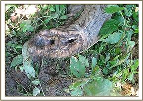
The male elephant calf was about one year old, the mother had sustained injuries on the left front leg and on the trunk. About two months ago the mother was rescued from a deep pit near Governors camp and later on it was found limping and lying in the thickets, it had a wound on the left front leg. It was then treated but the wound did not heal, the treatment was then repeated after a month but still there was no improvement. The trunk had been cut off by a snare about one year ago and it had adapted to using half the trunk for feeding and drinking. It had two young ones, a sub-adult and a juvenile but the older calf left and joined a larger herd when the mother could not move. The younger calf remained with the mother until the day it was rescued on 2nd April, 2008. The mother had been treated twice before it died.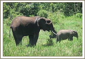
1st treatment
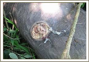
First treatment was done in February, 2008 about 200M from Little Governors Camp in a swampy area. The animal was in pain and had difficulties in walking. It was anaesthetized for treatment, the wound was deep and extending from medial towards the lateral side. The wound penetrated through the carpal joint. It was then debrided, drained and treated with antibiotics, antiflammatory drugs and multivitamins. It was then revived from anaesthesia and released though it was still limping and could not go far.

2nd treatment
One month later in March, 2008, the elephant had not improved, the limb was greatly swollen and could not move away from the swamp. It had started loosing much of its body condition. The veterinarian decided to repeat the treatment. The wound was again drained and flushed with hydrogen peroxide and a tincture of iodine, systemic antibiotics were also administered. A opening was also created using a surgical blade on the other side of the wound to enhance the drainage of pus and fluids from the wound. It was then revived from anaesthesia and thereafter closely monitored by the lodge managers who kept informing the veterinarian on its progress. The older calf had left the mother and joined the other herd.
On 1st April, 2008, the elephant was reported to have greatly deteriorated and emaciated, the calf had also lost much of its body condition and the veterinarian was informed to assist. It was found next to Little Governors camp on the Transmara side of the reserve. Due to continued infection on the limb, the animal had developed septiceamia and the other limb was getting swollen and the trunk developed infection at the tip and was exuding a lot of pus. It was therefore unable to walk or feed. There was agalactiae and calf was not getting enough milk and had grown weak. The elephant was completely unable to move and had poor prognosis and intense pain.
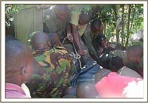
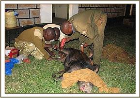
It was then anaesthetized and the wound examined and assessed but could not heal. A decision was then made to euthanize it and tusks recovered. The calf was then rescued by physical capture using a blindfold and ropes then later sedated using Azaperone hydrochloride (Stressnil) and transported by an aircraft to David Sheldrick’s Wildlife Trust (DSWT) orphanage in Nairobi. The calf was safely transported and released in one of the animal pens where it will be taken care of.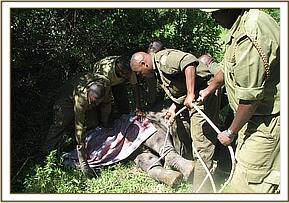

INVESTIGATION AND TREATMENT OF SICK LIONS IN MARA CONSERVANCY
Introduction:
On 4th April, 2008, two male lions in a pride of 6 males were reported to be showing signs of illness, they were lying down all day and not able to move or hunt. These cases were reported to Mara veterinary unit by the manager Mara conservancy. When the vet got to the ground, one of the sick lions was surrounded by other lions inside a thicket and could be seen lying and unable to move. The other lion was about 500meters away but in the same area. These lions were near Serena lodge/ Mara conservancy offices about 3kms away at a GPS location S -01.23.246, E – 035. 01. 183. It was not immediately known what condition it was but the most prominent clinical sign was hind-limb paralysis and panting with scanty saliva in the mouth.
Clinical observation
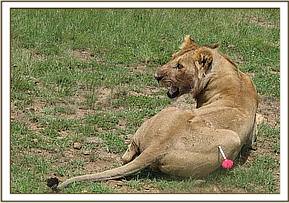
We decided to examine the first male lion which was isolated from the rest of the pride. Observations from a distant revealed hind-limb and front-limb paralysis, it was panting with wide open mouth that revealed scanty saliva in the mouth, very good body condition, unable to stand or walk, attempts to walk by crawling, hind-limbs and front-limbs unable to support the animal. The animal was very alert and grawling with a deep sound when approached from a vehicle. It had been fed on an impala the day before and the abdomen was distended. The head was always up-right with bright eyes and try to follow movements of vehicle and hear all the sound. It had been in an open area of hot sun for sometime when it was found.
Tentative diagnosis
Other sick lions were also reported to be having similar signs, observation from a distant and the clinical signs of paralysis could not reveal much for diagnosis, but the tentative diagnoses included the following:
- Rabies
- Canine distemper
- Viral induced myelopathies such as feline infectious virus (F.I.V)
- Nutritional deficiencies
- Tick paralysis
- Exposure to toxins
The three most likely diagnoses are, rabies, tick paralysis and poisoning. After consultations with other wildlife veterinarians and the team on the ground it was decided that one lion be anaesthetized, examined and later euthanized for postmortem examination and samples collected for laboratory analysis. This would then yield an accurate diagnosis that would help in treating other lions or controlling the condition from spreading to other prides.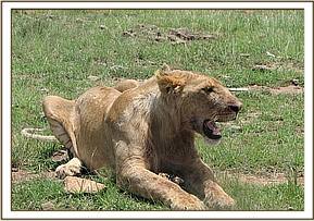

Chemical immobilization and capture
The lion was then darted from a close range using 35omgs of Ketamine hydrochloride combined with 350mgs of Xylazine hydrochloride and it took about 7 minutes to become fully anaesthetized. Then it was blindfolded and eyes were examined then treated with Opticlox eye ointment to avoid dessication.
Physical examination results
The vital physiological parameters were recorded as follows:
Respiration rate 24 cycles/ minute and deep.
Body temperature 38.3 degrees Celsius
Body weight about 150kgs
Pulse rate 72 beats / minute
Heart rate 72 beats / minute
Oral mucous membrane pink
Capillary refill time CRT 2 seconds
Decubital wounds on elbows and hock joint
Eyes normal
All the lymph nodes palpated and found normal
All the limbs and joints palpated and flexed but normal, no crepitation, no injuries, no swellings.
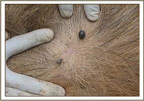
The skin and hair coat smooth but a number of ticks were found on the mane, groins and arm pits.
Blood samples collection
Blood was collected in 14mls plain tubes coated with clot retractor and in EDTA coated tubes. The blood was collected straight from the jugular vein and drawn into the tubes and kept in a cool box. No feacal sample was found from the rectum. Tissue samples from the ear was collected and kept in 95% ethanol for DNA analysis.
Euthanasia
The animal was euthanized using 40mls of 200% pentobarbitone hydrochloride (Euthatal) administered through the jugular vein. It then died after one minute and lifted to a safe place for postmortem examination.
Postmortem examination
The carcass was cut open from the neck to the pubic bone; all the thoracic and abdominal organs were examined in-situ before any manipulation. The lung, liver, kidney, urinary bladder, and lymph nodes appeared normal. Spleen was slightly enlarged but this could be due to the effect of euthatal. Intestinal lumen had haemorrhages and congestion of the walls with scanty intestinal contents. These organs were examined one after the other and pieces collected in zip-lock polythene papers and tightly sealed containers. The head was cut off and packed in a tight polythene bag and kept in a cool box full of ice-packs, the brain was to be tested for rabies infection. Other organs were kept in ice and in formalin for toxicology and histopathology tests. Stomach and intestinal contents were collected and frozen for toxicology tests. After collecting all the required samples, the carcass was then disposed far away from the reach of other lions and other animals.
These samples were then immediately flown to Nairobi -KWS lab the same day and later on submitted to the Government chemist for toxicology tests and veterinary laboratories for rabies test. No lab in Kenya could test serum for canine distemper. Histopathology samples were submitted to the University of Nairobi department of pathology for tests. Tick samples also taken to Kabete vet labs for identification to find out if they can cause tick paralysis. Some of the serum, blood and tissue samples remained in storage bank at KWS labs for follow up later.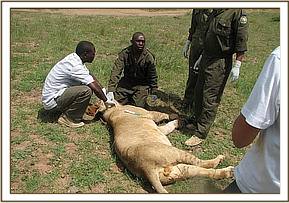

Examination and treatment of other lions
As the laboratory results were awaited two lions were recumbent and had similar signs as the first one, they were of the same age, same body weight and belonged to one pride. The veterinarian decided to give them symptomatic therapy as a final diagnosis awaited.
They were anaesthetized the same way as the first one and properly examined but the results remained same as the earlier one. Blood, tissue, feacal and tick samples were collected the same way and sent to KWS lab the same way.
The following treatments were instituted:
- Betamox long acting 40mls intramuscular injection
- Multivitamin 20mls intramuscular injection
- Ivomectin 0.2mgs/kg body weight (3mls) subcutaneously
- Fipronil (Frontline) application on tick infested areas
- Opticlox eye ointment applied on eyes.
- Dextrose solution infused intraveinously to help boost energy
- All the decubital wound sprayed using oxytetracycline spray
The animal was then revived from anaesthesia and fed on game meat since they were unable to hunt. One of the lions managed to recover and went but the latest one remained recumbent but had good signs of recovery.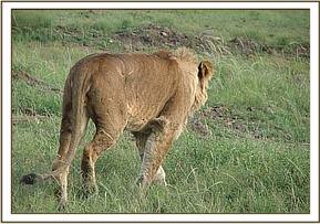

Laboratory results
The laboratory results so far received are shown in the table below, the rest of results are still awaited to help make a final conclusion on the cause of lion illness.
SAMPLE TYPE
TEST
LAB
RESULT
Stomach & intestinal contents
Toxicology
Government
Chemist
+ve carbamates (Furadan)
Liver,kidney,lungs,heart,LNs,spleen,
Intestinal tissues
Toxicology
Government chemist
+ve carbamates (Furadan)
Ticks for identification
Identification
Kabete vet labs
Not yet result
Brain tissue
Rabies (FAT test)
Kabete vet labs
Negative for rabies
Liver,kidney,lungs,heart,LNs,spleen,
intestinal tissues
Histopathology
University of Nairobi, Kabete
Not yet result
Serum
Biochemistry
KWS lab
Not yet result
EDTA blood
Haematology
KWS lab
Increased PCV, No haemoparasites
Discussion
The above results indicate that the cause of illness could have been carbofuran poisoning but more investigations need to be done and more tissue samples tested to confirm this.
Furadan is a trade name for Carbofuran which is a pesticide classified as an insecticide and nematocide. It belongs to the chemical family of carbofuran, the generic name being 2,3-dihydro-2,2dimethyl-7-benzofuranyl methylcarbamate. It was first registered for use as a pesticide in 1969 and is currently produced by FMC Corporation of U.S. Other trade names of this pesticide available in the market are Curaterr and Yaltox. It is used to control pests in fruits, field crops, vegetables, flower farms and forests. Methods of application are through aerial and ground application as a granular or spray form.
It acts by inhibiting the effects of Acetyl-cholinesterase enzyme that is involved in coordination of nerve functions, and with that it causes muscle paralysis and death of pests or any other animal in contact with it. Carbofuran (Furadan) is highly toxic to fish, birds and other wildlife species, it is known to leach through soil and has been found in groundwater as result of agricultural use.
These lions rarely go outside the park and how carbofuran chemicals got into the park is yet to be established. Serum biochemical tests should show the levels of cholinesterase enzyme as an alternative detection of carbofuran poisoning. The clinical signs were scanty and did not clearly indicate some of the signs of carbamate poisoning such as hypersalivation, cyanosis, vomiting and dyspnea but that does not rule out carbofuran poisoning if the intestinal contents, liver and other organs are tested and found positive for carbamate traces.
The lions had a history of feeding on a carcass of a hippo suspected to have died of the same poison. There are also reports of fish and other wildlife deaths along Mara river and all these cases are being investigated to know the problem. Lions are known to feed on fresh carcasses of other animals such as hippos and buffalos and if the death is due to poisoning then there are chances of lions getting poisoned. Mild poisoning can cause paralysis and prolonged suffering to animals before death; this seems to be the case with Mara lions.
Conclusion
Four lions in a pride of 6 showed signs of limb paralysis, one was euthanized to help with diagnosis, one was given symptomatic treatment and later recovered. The 3rd lion was treated but failed to recover and was later on found dead and suspected to have been killed by other lions when it was extremely weak, tissue samples were then collected and submitted to KWS lab by Mara conservancy management. The fourth lion was mildly affected and might have recovered. We have therefore lost two sub-adult male lions aged two and half years, others in the affected pride that have not been seen could have gone away or died.
Acknowledgement
We acknowledge the support of the management of Mara conservancy for assisting the KWS veterinarian in carrying out investigations and treatment on these lions. They helped in monitoring the sick lions and reported on their progress almost on a daily basis. They have also undertaken environmental audits to try find out the source of poisonous substance. Thanks to all those who participated in trying to save the life of Mara lions.
List of abbreviations
DNA - Deoxyribonucleic acid
EDTA -Ethyl-diamine tetra-acetate
CRT - Capillary refill time
FAT - Fluorescent antibody test
KWS - Kenya Wildlife Service
PCV - Packed cell volume
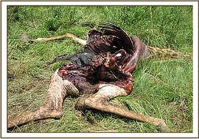
Postmortem examination of Giraffe that died in Mara:
The adult female giraffe was found dead just a few kilometers away from KWS research station. The carcass was about 3 days old and had been preyed on by lions, hyenas and vultures. There were bones of fetus around and it was suspected to have had dystocia (difficult calving).
Postmortem examination of a spotted hyena hit by a vehicle along Aitong road in Mara
This was a case of an adult male hyena that was found dead near Aitong town, the carcass was just a few hours old. Postmortem examination revealed fractured mandibles and canine teeth. The area residents said it had been knocked down by an over speeding vehicle at dawn. The carcass was then disposed of safely in the nearby thicket.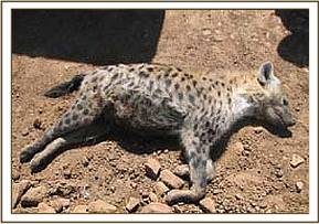

AFRICAN SWINE FEVER DISEASE SURVEILLANCE IN RUMA NATIONAL PARK:
Introduction
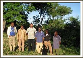
Kenya Wildlife Service (KWS) veterinary department in collaboration with International Livestock Research Institute (ILRI) conducted African swine fever (ASF) disease surveillance in Ruma National park during the month of April, 2008. The exercise went on for two weeks and involved capture and samples collection from Bush pigs within the park. Three veterinarians (two from KWS, one from ILRI), two capture rangers and a driver were involved in this activity.
This was the second time such an activity was taking place in Ruma, in November, 2007 two wild pigs were captured and blood, tissue and tick samples collected for laboratory analysis.
Highlights about ASF
African swine fever (ASF) is a highly contagious disease of pigs, first described in Kenya by Montgomery (1921). It is caused by a large DNA virus and is considered to be one of the most complex viral diseases to affect domestic animals. The disease is endemic in many African countries south of the Sahara desert. In Europe it is still endemic in Sardinia (Italy) and outbreaks were confirmed in the Alentejo region of Portugal in November 1999 (OIE, 1999). The disease has been successfully eradicated throughout the rest of Europe. ASF is classified in list A of the Office International des Epizooties (OIE, 1999) and inflicts significant socio-economic impact on affected countries.
Reservoir hosts and transmission of (ASF)
The ASF virus affects both wild and domesticated pigs and is transmitted by soft ticks of family Argasidae, genus Ornithodoros. In Africa, it is mainly transmitted through Ornithodoros moubata in Africa (Plowright et al., 1970) and Ornithodoros erraticus in the Iberian Peninsula (Sanchez Botija, 1963).
Ornithodoros corinaceus, a tick indigenous to the USA, has also been found to harbour and transmit ASF virus in experimental settings (Groocock et al., 1980), as has Ornithodoros savignyi which is present in Africa (Mellor and Wilkinson, 1985).
In Africa, ASF virus usually induces a non-apparent infection in three wild boar species: warthog (Phacochoerus aethiopicus), giant forest hog (Hylochoerus meinertzhageni) and bushpig (Potamochoerus porcus). Infection is characterized by low levels of virus in the tissues and low or undetectable levels of viremia. In the case of bush pigs P. porcus, viremia has been observed between 35 and 91 days following infection and the virus can persist in lymphatic tissues for 34 weeks (Anderson et al., 1998). Viral infection normally moves from these animals to domestic pigs through a biological vector, Ornithodoros moubata, and not by direct transmission.
ASF virus infections in the African vector Ornithodoros moubata are transmitted by transovarial and transtadial routes, whilst only transtadial transmission has been observed in the European vector Ornithodoros erraticus.
Significance and objectives of ASF surveillance
African Swine fever (ASF) is classified on OIE list A, which by definition contains only diseases that have the potential for very serious and rapid spread, producing serious socio-economic consequences (OIE, 1999). The last five years (1985-1990) of the ASF Spanish eradication programme cost approximately US $92 million.
Communities living around Ruma National park do keep pigs which are slaughtered occasionally for food especially during festivities. African swine fever is a disease of pigs but can induce a non-apparent infection in wild pigs such as bush pigs which then act as reservoir hosts for the disease and may transmit the virus to the domestic pigs.
The main objectives of ASF surveillance is to confirm if the bush pig population within the park have ever suffered from the disease by conducting serological tests to detect ASF antibodies in serum.
To test some of the diagnostic kits that have been developed for diagnosis of ASF in both domestic and wild pigs.
If the virus is found to be circulating in Ruma bush pigs then control measures can be put in place to avoid transmission of the disease to domestic pigs that can lead to an outbreak and serious economic losses due to pig mortalities in Kenya.
Methodology for bush pig capture and samples collection
Bush pigs are nocturnal animals that can only be seen late in the night, they are also extremely shy and cannot come close to humans. During day time they hide in deep thickets where they cannot be seen. These factors make it difficult to capture and handle bush pigs for sero-surveillance. In Ruma National park, they are commonly sighted near garbage collection sites within the residential areas at the park headquarters, Nyatoto gate, and Wiga gate. Apart from being in these areas, they are always seen at night while invading crop farms at night away from the park then hide in the nearby forest during day time. They mostly feed on gabbages and roots, they are also commonly reported as crop invaders at night.
Capture method and collaring
The use of nets for capture is the most reliable method and in Ruma, the capture team managed to capture one sub-adult male bush pig at night. The animals were trapped at night while grazing in an open space within the park headquarters’ compound, the escape routes were sealed by the net and when the animal tried to get into the bush it got entangled onto the net.
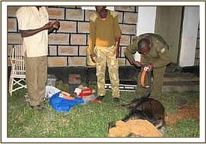
It was then physically restrained by hands and net. Later on it was tranquilized using Azaperone (Stressnil, 40mgs/ml concentration) a total dose of 20mgs was administered intramuscularly.
One of the captured wild pigs was fitted with a GPS collar that will help in monitoring their movements and collect more data on their interactions with domestic pigs in the area. This aspect will reveal the transmission trends of African swine fever disease between wild pigs and domestic pigs.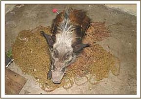

Samples collection
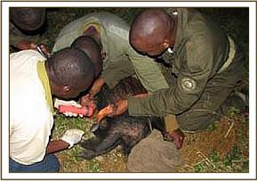
Blood samples were collected using vacutainer tubes and needles from the femoral vein then drawn into 14ml plain tubes coated with clot retractor and EDTA coated tubes and put in a cool box overnight. Ticks were picked from the skin of the animal using rat-toothed thumb forceps and placed in small containers and placed in a cool place. Tissue samples were also collected by cutting a small portion of the ear-pinnae. Hair samples also plucked out from the roots.
Samples storage and processing
The blood samples were centrifuged at 1500 rounds per minute for 10 minutes then serum extracted into cryovials and kept in a deep freezer for submission to the lab for analysis. Tick samples were also kept a deep freezer, tissue samples were dissolved and kept in a freezer too.
Prevention and Control of (ASF)
Control of the disease is usually achieved by the control of tick vectors. No treatment or vaccine is available for ASF and control of this disease is based on rapid laboratory diagnosis and the enforcement of strict sanitary measures.
Depending on the epidemiological status of the disease in a particular region, different measures are recommended. In endemic areas of Africa, the most important factor is to control the natural tick vectors and wild pig reservoirs, and/or limit their contact with domestic pigs. The identification and slaughter of sick and carrier animals is crucial to the control of the disease.
Conclusions/Recommendations
The work was concluded after two weeks, the intention was to capture more than 10 bush pigs but due to low numbers of bush pigs in the park and their unpredictable movement patterns and difficulties of capturing at night, the team managed to capture and collect samples from one male bush pig. The recommendations for the future work were;
· Put on collars to any bush pig captured for ease of tracking and location for continuous monitoring and samples collection over a period of time.
· The duration of biological samples collection for this project can be staggered over a longer period of time probably one year because it is only possible to capture 2 or 3 at a given time then they disappear.
· Opportunistically capture bushpigs whenever vet team goes to Ruma to attend to other cases and collect the required samples.
· Use of baited traps well designed for capture of bushpigs.
Conclusion
It had been a rainy season in Mara and other parts of Central rift, most animals have moved out of the reserve into community areas and animals have plenty of grass and water within the reserve. Most areas are not accessible due to bad weather roads and other than carbamate toxicity in the lions of Transmara, there were no many reported cases during the month. There are several ongoing research activities in Mara which the veterinary unit is actively involved in, these include wildlife diseases investigations such as mange infestation in cheetahs, cheetah monitoring and conservation project, elephant monitoring activities, investigations on carbamate toxicity in lions.
Reported by; Dr. Domnic Mijele