EASTERN CONSERVATION AREA VETERINARY UNIT MONTHLY REPORT APRIL 2017 Report by: Bernard Rono Summary This report describes the activities of the Meru Veterinary Unit in Northern Kenya in April 2017
EASTERN CONSERVATION AREA VETERINARY UNIT MONTHLY REPORT APRIL 2017
Report by: Bernard Rono
Summary
This report describes the activities of the Meru Veterinary Unit in Northern Kenya in April 2017. This month marked the onset of long rains in Samburu and Isiolo areas; however other areas such as Laikipia and Meru Conservation area received inadequate rainfall. There was insufficient pasture in these areas and livestock incursion into conservation areas continued throughout the month.
The Vet Unit attended to an investigation into the cause of death in the impala population in Samburu National Reserve, a white rhino showing lameness and a post mortem examination on an elephant which died of natural causes in Meru national park.
The Meru veterinary unit is supported by the David Sheldrick Wildlife Trust to provide veterinary care in northern Kenya.
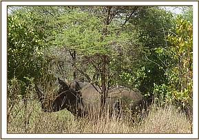
CASE #1: LAMENESS IN A WHITE RHINO
Date: 11th April 2017
Species: White Rhino
Sex: Male
Age: Adult
Location: Meru national park
History
The rhino monitoring team in Meru National Park reported a white rhino which showed lameness on its left foreleg. Close observation showed this rhino was in good body condition and there were no visible injuries. No treatment was required.
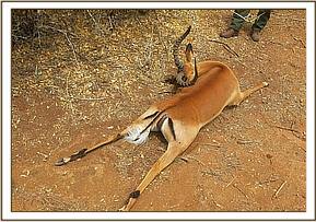
CASE # 2: IMPALA MORTALITY IN SAMBURU NATIONAL RESERVE
Date: 18th April 2017
Species: Impala
Location: Samburu National Reserve
History
The Meru Veterinary Unit responded to a call by the game warden in charge of Samburu National Reserve to investigate the cause of death in the impala population within the reserve. Three fresh impala carcasses were found on the 16/03/17, this number rose to six on 18/03/17. Only one breeding herd of impala was affected near the Larssen’s camp. Carcasses consisted of four (4) adult female, one (1) male and one (1) sub-adult male impala.
We conducted a post mortem examination on one carcass and collected samples for laboratory analysis.
General observations
- The deaths coincided with the onset of the long rains in the Samburu area two weeks earlier. Hence vegetation in the reserve was rejuvenated with plenty of lush grass.
- The deaths occurred in only one breeding herd of impalas consisting of approximately 40 individuals, no other species was affected. One live female impala in the herd appeared weak although it was in good body condition
The carcasses were generally in good body condition although they were bloated due to high ambient temperature and rapid putrefaction as a result. Some carcasses were partly scavenged and internal organs were missing while others were infested with various stages of maggots. Blood tinged bodily fluids oozed from orifices due to putrefaction.
Post mortem examination
We conducted a post mortem examination on an apparently fresh but bloated male carcass.
- The prescapular lymph nodes were enlarged.
- Gastrointestinal tract contained plenty of ingesta and undigested grass material.
- Spleen and liver were normal. The kidney was dark and autolysed.
Samples collected at post mortem were as follows:
- Impression smears of the prescapular lymph nodes and muscle tissue and samples of lymph nodes preserved separately in 10% formalin and ethanol
- Skeletal muscle tissue, liver and spleen preserved in 10% formalin and frozen for toxicological analysis
Diagnosis and management
Tentative diagnosis was clostridium enterotoxaemia caused by Clostridium perfringens Type D also known as pulpy kidney. In addition the to post mortem picture (autolysed kidney) and environmental conditions, smears revealed gram positive rods identified as Clostridium spp. Confirmatory diagnosis requires demonstration of toxins from intestinal filtrates and subsequent identification by neutralisation with specific antiserum.
C. perfringens form part of normal flora in the gastrointestinal tract (GIT). However in presence of certain factors these bacteria can cause disease. Predisposing factors for this disease include altered GIT environment for instance due to ingestion of vegetation which promote excessive growth of bacteria and production of toxins. The toxins produced cause vascular damage particularly in the brain.
In wildlife under free range management the disease is considered self – limiting and coincides with the onset of the rains after a prolonged dry period. No further action was taken on the affected impala herd.
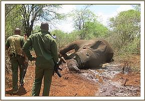
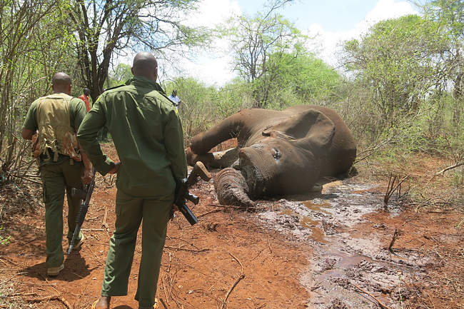
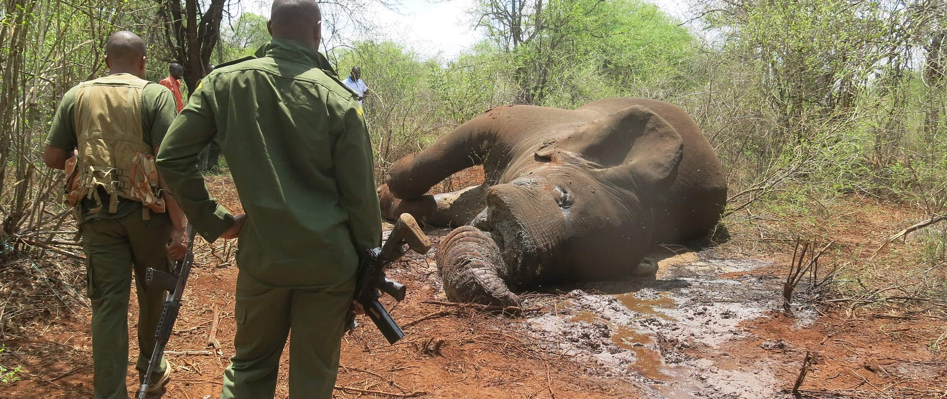
CASE #3: POST MORTEM EXAMINATION OF ELEPHANT IN MERU NATIONAL PARK
Date: 21st April 2017
Species: Elephant
Sex: Male
Age: Adult
Location: Meru National Park
History
The Wildlife Monitoring team in the Rhino Sanctuary reported an elephant carcass during routine patrol. Both tusks were intact and removed by the patrol team for safe keeping. The carcass was extensively autolysed and heavily infested with maggots. The Perineum was partly eaten by scavengers and internal examination of the carcass was not feasible because of its state of decay, however, death was attributed to natural causes.