EASTERN CONSERVATION AREA VETERINARY UNIT MONTHLY REPORT JUNE 2017 Report by: Bernard Rono Summary This report describes the activities of the Meru Veterinary Unit operating in Northern Kenya in June 2017
EASTERN CONSERVATION AREA VETERINARY UNIT MONTHLY REPORT JUNE 2017
Report by: Bernard Rono
Summary
This report describes the activities of the Meru Veterinary Unit operating in Northern Kenya in June 2017. Rainfall patterns in northern Kenya were erratic with little or no rainfall recorded in some parts.
During this month we attended to bone fractures in a giraffe and an elephant. An elephant which strayed into community land in Machakos county was captured and relocated to Mwea National Reserve. In Lewa Conservancy a white rhino was treated for lameness caused by a fight and in Ol Pejeta Conservancy a post mortem examination was carried out on a black rhino carcass to determine the cause of death.
We acknowledge the David Sheldrick Wildlife Trust for financial and logistical support for the Meru veterinary unit operations in northern Kenya.
CASE#1 FRACTURE IN A GIRAFFE
Date: 4th June 2017
Species: Reticulated giraffe
Sex: Male
Age: 6 months
Location: Loisaba Conservancy
History and Mangement
This young male had an open complete fracture of the right metatarsal bones midway between the hock joint and fetlock joint. The foot was attached by a piece of skin and was dragging as the animal walked causing alot of pain.
The only option for this animal was euthanasia to relieve it of further suffering.
Autopsy findings
The carcass was in good nutritional condition with a smooth shiny hair coat.
The soft tissue around the fracture site was decomposed and the foot was decomposed with the hoof loosely attached. There was an area of congestion on the subcutis of the right flank over the 7th and 8th rib possibly following a heavy fall at the time of the fracture.
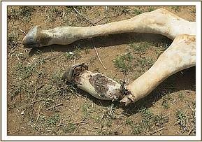
All internal organs were normal and there were no signs that the infection on the fracture site had spread systemically.
CASE#2 POST MORTEM EXAMINATION ON A BLACK RHINO CARCASS
Date: 5th June 2017
Species: Black Rhino
Sex: Male
Age: 5 years
Location: Ol Pejeta conservancy
History
This rhino was initially treated on May 16, 2017 for injuries to the back muscles and left hock joint sustained following an attack by lions. Despite the treatment it was reported that its body condition continued to deteriorate and there were signs of infection in the wounds. On June 6, it was found dead in a river with the front and rear horns intact.
A post mortem examination of the carcass was carried out to record the cause of death. Observation showed rupture of the left calcaneal (achilles) tendon, ligament tear and instability of the bones of the hock joint. There was a septic bite wound to the vertebral muscles dorsal to the sacrum, this extended deep into the sacral bone.
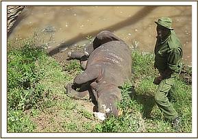

On opening the carcass there was froth and ingesta on the trachea and the bronchi, this indicated aspiration prior to death. The lungs were normal and floated freely when immersed in water. There were no other significant findings in the internal organs. These findings are consistent with asphyxiation due to drowning.
CASE#3 TREATMENT OF WOUNDS IN A WHITE RHINO
Date: 07/06/17
Species: White rhino
Sex: Male
Age: 8 years
Location: Lewa Wildlife Conservancy
History
The white rhino had fight wounds spread generally across the whole body with the most significant one being on the left forelimb close to the middle toe. The foot was swollen and as a result the rhino presented with weight shifting lameness.


The rhino was immobilized using 6mg of Etorphine hydrochloride and 50mg of Azaperone combined in one intramuscular dart. After the rhino went down, a blind fold and ear plugs were applied and anesthesia stabilized using 10mg Butorphanol tartarate administered intravenously at the ear vein. Ventilation was aided using a mechanical ventilator and vital parameters monitored after 5 minute intervals. Body temperature was kept down by pouring cold water on the animal.

The wounds were all cleaned and disinfected using Povidone Iodine soaked swabs and Hydogen peroxide. The rhino was treated with 2500mg Flunixine meglumine, 15000mg Amoxicillin trihydate and 100mg of Cyanocobalamine. The wounds were then sprayed with Oxytetracycline aerosol.
Reversal and Prognosis
The anesthesia was reversed by administration of 100mg of Naltrexone hydrochloride, (50mg I.V and 50mg I.M). The rhino is expected to make a complete recovery in the coming days.

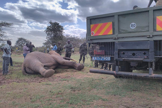
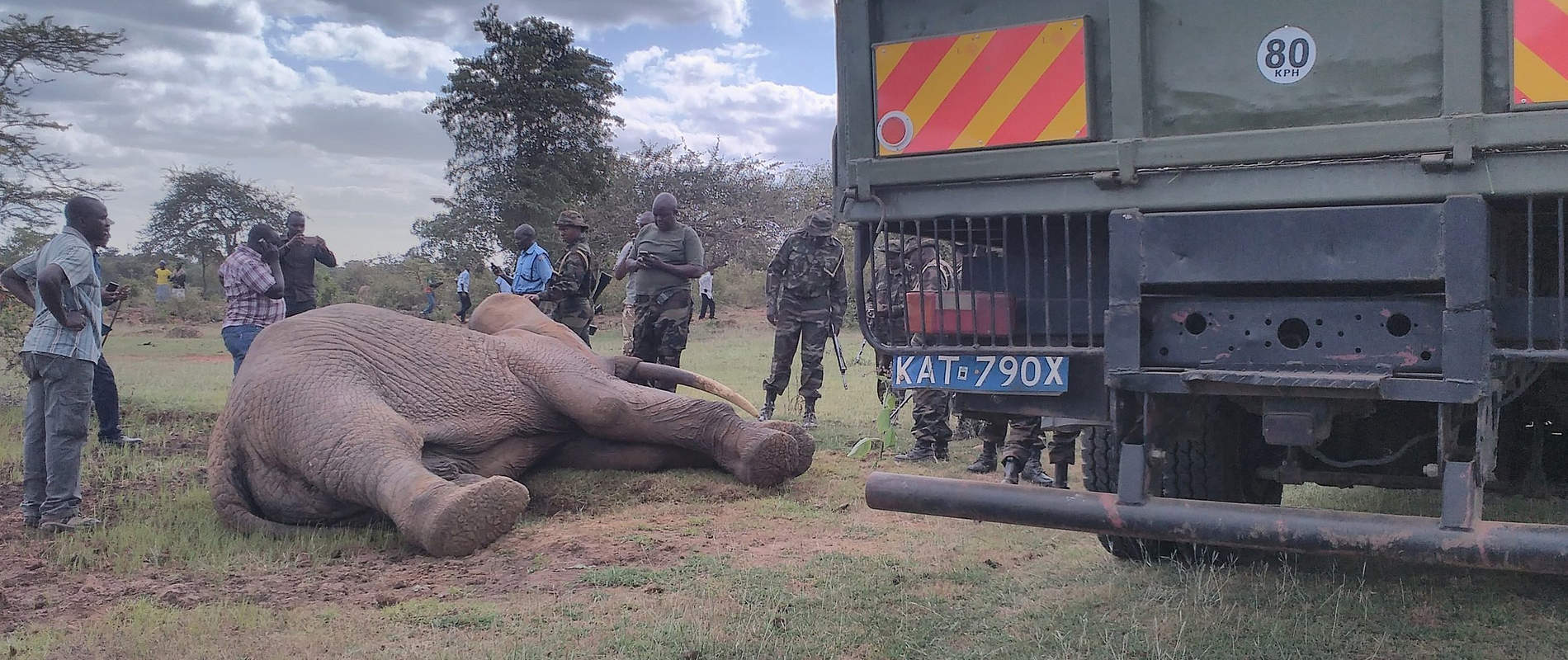
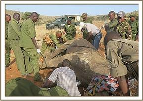
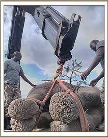
CASE #4: RELOCATION OF A STRAY ELEPHANT
Date: 11th June 2017
Species: Elephant
Sex: Male
Age: 30 years
Capture site: Matangi, Machakos
Release site: Mwea national reserve
History
On the 10th June, the KWS warden in Mutomo, Kitui County reported that an elephant had strayed from Mwea National Reserve and had been wandering in villages on the boundary of Kitui and Machakos County for the past week. Residents reported crop damage and risk of human and livestock injury or loss of life. Teams from Mwea, Mutomo and Ol Donyo Sabuk stations had attempted to drive the animal back but were not successful.
This elephant was captured, transported by road and released in Mwea National Reserve on 11/06/17.
Capture and loading into the truck
The elephant was located in a thicket which was not accessible by road. The vet darted the problem elephant from a helicopter using 20mg Etorphine hydrochloride 20mg in a 3ml dart. After darting it was driven to open ground before the drugs took effect four minutes later.
The trunk was straightened to ensure a patent airway during loading while its ear flap was used to cover its eyes. To keep the temperature within normal limit the animal was doused with water regularly during loading and transportation.
Physical examination showed superficial wounds inflicted by a sharp object on its trunk and shoulder. The wounds were washed and infused with Povidone iodine. An antibiotic and Catosal, a multivitamin were given intramuscularly.
The elephant was hoisted onto a flat-bed truck by a crane and restrained using ropes on lateral recumbence. The truck and escort team departed for Mwea NR at 1553 hours.
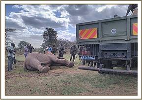

Transportation and release
This elephant was transported under deep sedation which was maintained using additional doses of Etorphine hydrochloride administered intravenously through the superficial ear vein. Respiration and reflexes of the ear flaps were monitored during transportation for signs of recovery. Etorphine, 5 mg, was given two hours after the initial dart and subsequent maintenance doses at an hourly rate of 5mg until release. In total 35mg Etorphine dose was administered for induction (20mg) and maintenance (15mg).
The distance covered by road was 120 kilometers and total travel time was 3 hours and fifteen minutes from capture site to release site. A suitable site in the middle of the park was selected for release not far from water point.
Reversal
The anesthetic was reversed using 30mg Diprenophine hydrochloride. Three minutes later the elephant was fully reversed and in standing position. Initially the elephant showed a staggering gait due to numbness of the hind legs but it slowly recovered and walked away.

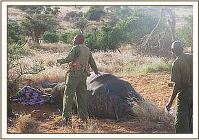
CASE #5: LAMENESS IN AN ELEPHANT
Date: 14th June 2017
Species: Elephant
Sex: Male
Age: Adult
Location: Lesiamo, Ndoto ranges
History
Wildlife scouts from the Milgis Trust reported that an elephant in the Ndoto mountains showed lameness and dragging of it front leg for the past ten days. They requested veterinary examination to determine the cause of lameness. This elephant was tracked down and darted on 14/06/17.
Immobilization, examination and management
The vet immobilized the animal from foot using Etorphine hydrochloride delivered in a 3.0 ml dart. The dart was placed in the neck muscles and the elephant was sedated after 4 minutes on sternal recumbence. For physical examination the elephant was tipped to left lateral recumbence.
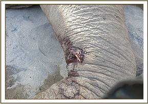
The left forelimb was swollen distal to the elbow joint. It had an open wound which was discharging pus. There was a complete open fracture of the ulna bone with bone fragments protruding from the open wound. The cause of the fracture could not be established immediately.
Although this elephant was in good body condition, this fracture compromised its health and wellbeing. Fractures in large herbivores are difficult to repair due to its weight and inability to fix the affected bones and restrict movement. The fracture site was also septic and there were chances of infection spread to other parts of the body.
This elephant was euthanized to relieve it of pain and suffering attributed to the fracture.