FIELD VETERINARY REPORT FOR MASAI MARA - FEBRUARY 2015 Reported by Dr
FIELD VETERINARY REPORT FOR MASAI MARA - FEBRUARY 2015
Reported by Dr.Campaign K. Limo
Introduction
The month was characterized by gradual increase in precipitation in the beginning which reduced and finally stopped towards the end. Many elephants were seen in the reserve. A few cases of human inflicted injuries on animals were however recorded and attended to. Below are cases attended to during the month.
CASE#1 COLLARING AN AARDVARK FOR RESEARCH PURPOSES
Date: 7th Feb2015
Species: Aardvark
Age: Adult (35-40yrs)
Sex: Female
Location: Talek Area
Date: 7th Feb 2015
History
Though prevalent in most conservation and pastoral areas little is known about this nocturnal mammal. Aardvark activities have not been recorded in these areas of late possibly because of a change in land use. Little research has been carried out on this species.
KWS in collaboration with other researchers sponsored by Bridge Earth Limited Japan carried out the collaring of one aardvark in Masai Mara.
Aims:
- Study effects of human activity on aardvark activity
- Get baseline information on aardvark social structuring, habitat use and ranging patterns
- Promote an understanding of aardvark conservation among international and local stakeholders
- Produce a documentary on aardvark
Capture and collaring
After several unsuccessful attempts to capture this animal using strategically placed capture nets, the capture team decided to actively search for these animals at night using good lights and dart them. A combination of 2mgs Medetomidine and 160mgs Ketamine in a 3ml daninject dart was used to immobilize a female spotted at 3am in the morning. She was prevented from accessing any burrow by people and vehicles before the drugs took full effect. The drugs took full effect after eight minutes as she tried to dig a new burrow.
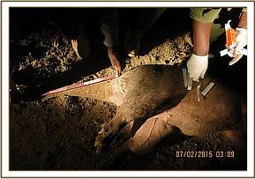

After making sure she was stable, a light satellite collar was deployed and tissue and hair samples were collected. The animal was given intramuscularly 1500mgs Amoxicillin antibiotics and 14mgs Dexamethasone Sodium anti-inflammatory to prevent shock.
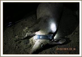

Reversal
The aardvark was revived by intramuscular administration of 9mgs Atepamezole one hour after immobilization. She woke up after four minutes and tried to pull off the collar before calming down and moving on. A team is monitoring her and her signals are being received at night when she comes out.
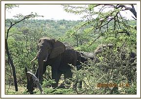
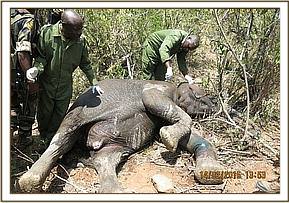
CASE#2 TREATMENT OF A LAME ELEPHANT BULL
Date: 9th Feb 2015
Species: African elephant
Age: Adult (35-40yrs)
Sex: Male
Location: Naboisho conservancy
History
This massive bull was seen by Naboisho conservancy rangers showing lameness on one of his forelimbs. He was in a herd of about twenty elephants but kept lagging behind.
The elephant was in a rocky valley making it difficult to access and immobilize in such a terrain. The other members of the herd moved out when they felt the team’s presence but he remained. The carpal joint was swollen and he could hardly place weight on this limb. He finally left the rocky valley and entered an open area suitable for darting.

Immobilization examination and treatment
Immobilization was achieved by darting him with 16mgs Etorphine delivered through a 3ml Daninject dart from a vehicle. The drugs took effect after seven minutes with the elephant going down on his left side. Examination revealed a swollen right carpal joint with no visible wound. Aspiration of the swelling yielded dark blood. The joint and the bones around this area appeared intact and the swelling was suspected to have been caused by a sprain to the joint.
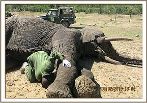

This elephant was given 15000mgs Amoxicillin antibiotics and 3000mgs Flunixin meglumine anti-inflammatory intramuscularly. The joint was also massaged for ten minutes to try and improve circulation.
Reversal
The elephant was revived by administration of 48mgs Diprenorphine hydrochloride through the ear vein. The elephant woke up to join other members of the herd after three minutes.

Prognosis
Good. This elephant was seen two weeks after treatment having walked a distance of about 40km in much better state. He will still be monitored and treated again when necessary.
CASE#3 EXAMINATION OF TWO BULL ELEPHANTS IN SIANA CONSERVANCY
Date: 9th and 11th Feb 2015
Species: African elephant
Age: Adult (35-40yrs)
Sex: Male
Location: Siana conservancy
History
These two bull elephants in Siana conservancy were reported with injuries by Siana Conservancy management. According to the report, rangers had seen one bull limping with a swelling on one of his forelimbs and the other with a wound to the right side of his withers.
Examination
On the 9th Feb 2015 a search for the elephants was fruitless. They were said to be in an inaccessible thicket as only their tracks could be seen and the search was called off after dark.
On the 11th Feb 2015 the two bulls were spotted close to the border of Siana and Olarro Conservancies and management contacted the vet team for assistance. The elephants were found in a small thicket and appeared elusive. One was seen as he emerged and then he dashed back into the thicket. He appeared to walk with ease and no injury was noted. The second elephant was observed from all sides and no injury was seen. The management will keep monitoring the area and will broaden their search as it is likely these were not the two reported injured elephants.

CASE#4 REPLACEMENT OF AN ELEPHANT’S SATELLITE COLLAR
Date: 12th Feb 2015
Species: African elephant
Age: Adult
Sex: Female
Location: Naboisho conservancy.
History
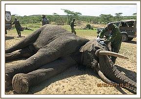
This female adult named Caroline was collared in 2011 and the lifespan of the GSM-Satellite collar that had been deployed was coming to an end. The collar has been used to monitor this elephant and its herd’s movements which has helped advise management on the conservation and protection of these animals. There was therefore need to replace this collar before its signal completely disappeared and this was done together with the Mara Elephant Project and Save the Elephant teams. Caroline was found with other members of her family; a four year old calf, a sub-adult bull and an adult female. She appeared agitated on approach possibly due to prior experience of immobilization.
Immobilization and collaring
Caroline was immobilized by use of 16mg Etorphine hydrochloride delivered through a 3ml dan inject dart from a vehicle. The drugs took full effect after seven minutes by which time she had moved about a 100 meters with the other family members and she went down on her right lateral.
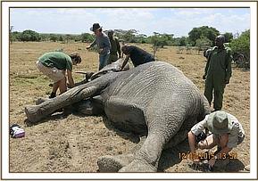

The other family members were protective of her but were scared away by vehicles to give room for the collaring procedure. After making sure she was stable, the old collar was removed and the new one fitted. Soundness of the collar was confirmed before the elephant was revived. This immobilization also afforded the team a chance to examine Caroline for any health issues. She appeared to be in good condition, except for few scratches and abrasions common with elephants in the wild. She was given 15000mgs Amoxicillin antibiotics and injectable multivitamins intramuscularly.
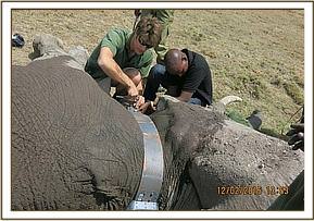

Reversal
Caroline was revived by administration of 48mgs Diprenorphine hydrochloride through the superficial ear vein. She woke up in three minutes and joined the rest of the family waiting nearby.

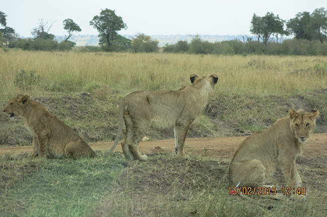
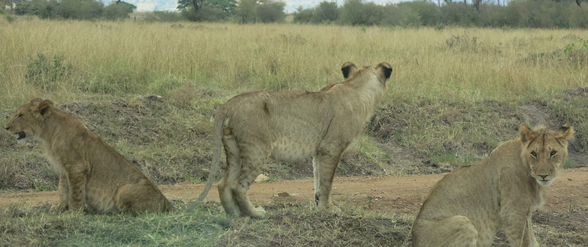
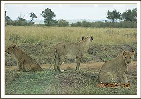
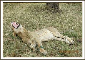
CASE#5 TREATMENT OF AN INJURED LION
Date: 13th Feb 2015
Species: Lion
Age: Sub adult
Sex: Male
Location: Mara Triangle
History
This young male was seen by Mara Triangle rangers limping on his left forelimb. The team found him under a tree near the road with other members of the pride as well as three young cubs. He stood as the vehicle approached allowing the vet to examine the injury. The left forelimb at the carpal joint was severely swollen and was not weight bearing.
Immobilization, examination and treatment
Immobilization was achieved by a combination of 2.5mgs Medetomidine and 160mgs Ketamine delivered through a 3ml daninject dart from a vehicle. The drugs took effect after eight minutes and a blindfold was applied and the lion placed in comfortable position. Cloxacillin ointment was applied to both eyes to prevent desiccation.

Closer examination of the affected limb revealed a deep wound to the lateral surface of his left carpus caused by a bite, most likely from another lion. The wound was becoming septic and was cleaned with Hydrogen peroxide and swabs. Copious amount of water was used to rinse before tincture of iodine was used to disinfect the wound. Oxytetracycline spray was applied topically and then 1500mgs Amoxicillin antibiotic and 12mgs Dexamethasone Sodium anti-inflammatory were administered intramuscularly.
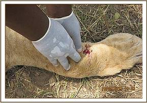

Reversal
The lion was revived by intramuscular administration of 10mgs Atepamezole hydrochloride one hour after immobilization. He woke up after eight minutes and called for other pride members who came to join him.

Prognosis
Good.
CASE#6 TREATMENT OF AN ELEPHANT WITH AN ARROW WOUND
Date: 14th Feb 2015
Age: 8 years
Species: African elephant
Sex: Male
Location: Olarro Conservancy
History.
This young elephant was seen isolated in a bush by the Olarro conservancy rangers. The vet team found him in a thicket close to a watering point, elusive and agitated. His left forelimb was swollen at the carpus and he moved with difficulty.
Immobilization, examination and treatment
Immobilization was achieved by use of 8mgs Etorphine hydrochloride delivered through a 1.5ml Dan inject dart from foot. It took eight minutes for the drugs to take full effect with the elephant assuming left lateral recumbency.
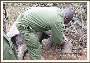

Examination revealed a moderately swollen left carpus with a small penetrating wound to the medial surface of the joint, most likely caused by an arrow. Further probing of the wound yielded no foreign body but the wound was becoming septic. However, the joint integrity was not affected. The wound was washed with copious amount of water, Hydrogen peroxide and tincture of iodine to clean and disinfect. Green clay was then packed into the wound to promote faster healind. Finally, 15000mgs Amoxicillin antibiotics and 1500mgs Flunixin meglumine anti-inflammatory, were given intramuscularly.

Reversal
The elephant was revived by administration of 18mgs Diprenorphine hydrochloride through the superficial ear vein. He woke up in three minutes and briefly tried to charge at the attending team before retreating deeper into the bush.

Prognosis
This elephant is expected to make a full recovery.
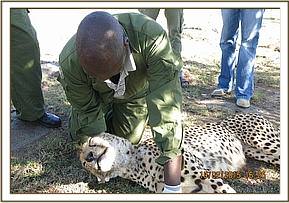
CASE#7 POST MORTEM OF A CHEETAH
Date: 19th Feb 2015
Species: Cheetah
Sex: Male
Age: 2 years
Location: Olare Orok conservancy
History
This cheetah’s brother had been found dead on the 18th Feb 2015 and there were concerns over his health as well. He was found dead in the afternoon after frantic efforts to search and treat him.
The cheetahs were seen with teary red eyes and nasal discharge by tour guides who reported the incident to The Mara cheetah project team. Their cheeks also appeared swollen and they were having difficulty breathing. Two other cheetahs, a mother and daughter, had been seen a month earlier with similar symptoms, 20km from the two latest cases. However, they were constantly monitored and the swellings subsided and they made a full recovery.
Another case of an adult female had also been reported two months ago by the Meru Mara cheetah project team. However, the swelling resolved in a week and the cheetah also made a full recovery. The two latest cases unfortunately succumbed. Both carcasses were brought together and a representative post mortem was done on the fresher of the two carcasses.
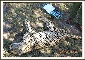

General observation
- On estimation, the carcass was less than twelve hours old
- The carcass appeared to have been in good body condition before death
- No physical injuries were evident on the body
- The head appeared swollen down to the midneck
- Eyes were swollen with greenish discharge and the eyelids appeared glued together
- There was some colourless discharge from the nostrils
- Rigor mortis was present
- Oral mucocutaneous junctions appeared ulcerated

On opening the carcass, the following findings were noted:
- The neck was severely swollen with signs of gelatinous fatty degeneration of tissues around the neck and surrounding areas. This degeneration was also evident to some extent in other areas of the body
- The lymph nodes around the head and neck were swollen, especially the retropharyngeal lymph nodes and parotids
- There were petechial haemorrhages which were more intense on tissues around the thorax cranially towards the head. There was less petechiation on more caudal tissues
- Lymphadenopathy appeared localized with degree of abcessation forming near the retropharyngeal lymph nodes
- Pericardial sac had slightly viscous fluid otherwise the heart appeared normal with slight petechiae
- There were signs of pleurisy with more than expected fluid seen in the pleural cavity
- Spleen appeared normal in size and consistency. It was flaccid
- The liver and both kidneys appeared congested and slightly darkened
- Though they appeared normal in size, the lungs had whitish dots, diffusely distributed thought to be complex deposition on its parenchyma. Some mucous were also found at lower levels of respiratory tree
- The stomach and the entire lower gastro intestinal tract were virtually empty suggesting that this cheetah had not fed for a couple of days. Tapeworms were found stagnating in the lumen of the larger intestines
- All other organs appeared normal
Blood smear for microscopy, skin tissue samples for DNA and lung, liver and kidney tissues for histopathology were collected.
Conclusion
Based on history, clinical and post mortem picture, the signs are consistent with those of cat flu viral infection with its associated complications. Cheetahs with good immunity can make a full recovery. Other factors including genetics and immunity status could have compromised ability of these cheetahs to fight this infection. Samples collected will be used to confirm the diagnosis.
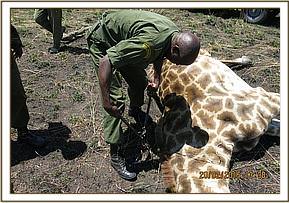
CASE#8 DE-SNARING A GIRAFFE
Date: 20th Feb 2015
Species: Masai giraffe
Sex: Female
Age: Adult
Location: Mara triangle
History
This snared giraffe was reported by the Mara Triangle Conservancy rangers. She was seen coming from the escarpment bordering the conservancy with a snare on the neck and dragging a tree stump along.
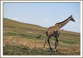

Immobilization, examination and treatment
Immobilization was achieved by administration of a combination of 12mgs Etorphine hydrochloride and 50mgs Azaperone delivered through a 3ml daninject dart. The drugs took effect after eight minutes with the giraffe falling down and assuming right lateral recumbency. The braided winch wire snare was cut loose and pulled out. Though there were no injuries seen, the giraffe was given prophylactic antibiotic treatment of 6000mgs Oxytetracycline and 40mgs dexamethasone anti-inflammatory intramuscularly.

Reversal
The giraffe was revived by administration of 24mgs Diprenorphine hydrochloride intravenously through the jugular vein. The giraffe rose almost immediately and walked away maintaining a left lateral kink of the neck posture. This was caused by prolonged dragging of the tree stump along with the snare. She was manually restrained with ropes and the neck massaged in an attempt to straighten it. The neck straightened for a few minutes but upon release soon resumed to its kink posture.
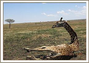

Prognosis
Guarded to fair. The kinked neck might interfere with ease of movement making this giraffe an easier target for predators. However the neck is expected to straighten with time. Mara conservancy rangers were advised to keep watch on this giraffe and report the development.
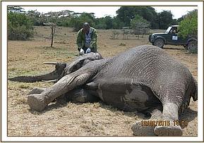
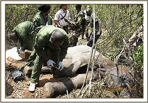
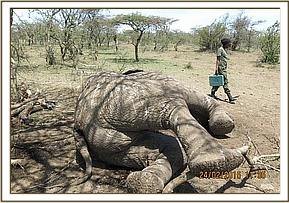
CASE#9 POST MORTEM OF A SPEARED ELEPHANT
Date: 24th Feb 2015
Species: African elephant
Sex: Male
Age: Adult (35-40yrs)
Location: Naboisho conservancy
History
This elephant carcass was seen by the Naboisho conservancy rangers, Mara elephant project rangers and KWS rangers on patrol. The tusks were intact.
General examination of the carcass
- Carcass was relatively fresh, about three days old
- The elephant appeared to have been in good health before death with a score of 4 out of 5
- There was no evidence of struggle before death
- Scavengers had begun eating parts of the body.
- There was a penetrating wound to the left flank whose margins had been extended by scavengers clearly caused by sharp object most likely spear
- On further examination, the penetrating wound was deep enough and involved the peritoneum. The object had punctured the stomach and large intestines causing peritoneal contamination.
Conclusion
This elephant succumbed to acute peritonitis sequel to trauma. Spearing was the most likely cause of trauma. Both tusks were retrieved and handed over to security officers for accounting and safe custody.

Acknowledgements
Mara veterinary unit would like to extend their gratitude to all members of the public who assisted in spotting, reporting and monitoring of animals that required help during the month. Thanks to management of various conservancies who keep vigil and subsequently report cases which require intervention. Thanks to the Mara cheetah project and Mara Meru cheetah project teams who kept constant vigil on the cheetahs and provided updates on their status to the unit. Thanks too to Minara foundation through The David Sheldrick Wildlife Trust and Kenya Wildlife Service for their immense support to the unit.
Together we can make a difference in saving the very unique heritage within our territory.