The Mt Kenya Wildlife Veterinary unit attended and treated several wildlife cases that required urgent veterinary intervention in the whole of Laikipia-Samburu ecosystem and Mt. Kenya region during the month of November, 2018.
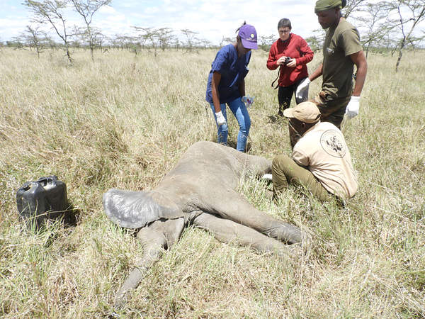
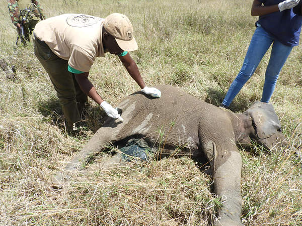
Case #1: Treatment of an injured elephant calf
Animal Identification
Species: African Elephant (Loxodanta africana)
Sex: Male
Age: Juvenile
Location: Loisaba Wildlife Conservancy
Date of clinical intervention: 3/11/2018
History: A juvenile male elephant was sighted in Loisaba Wildlife Conservancy limping and in a lot of pain
accompanied by its mother and another sub-adult elephant .
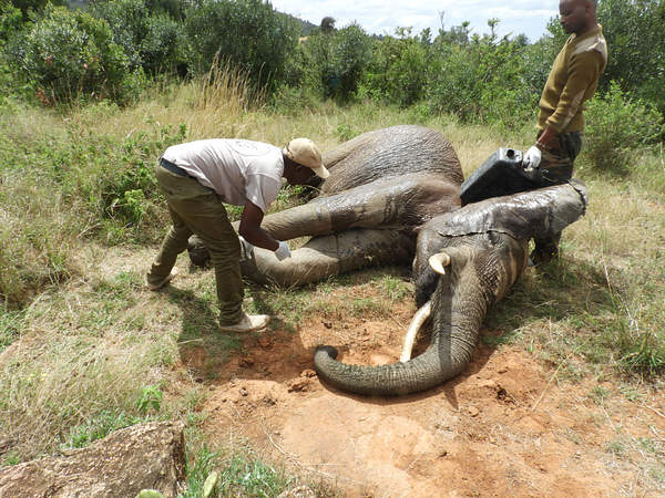
Immobilization, Examination and Treatment: The injured elephant was found in a fairly thick area and was darted from a vehicle using 5mgs of etorphine Hcl in a 1.5ml Dan-inject dart. It got adequately immobilized after about 5 minutes and went down on lateral recumbency, The mother refused to leave the calf despite making several attempts to push it away and was therefore darted using 17mgs of etorphine Hcl.
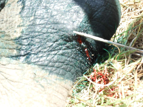
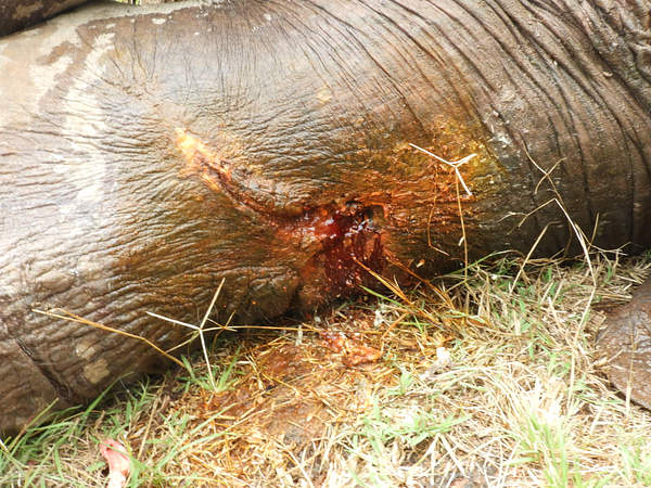
The elephant had one deep penetrating wound extending posterior-anterior and exit on the front side. The wound was already infected with a lot of necrotic debris and exudates. It was a bullet wound that was shot from behind, hitting the leg and exiting, leaving a deep narrow wound through the leg. The affected leg was heavily swollen and painful.

The wound was thoroughly cleaned with lots of clean water then probed using sterile gauze swabs attached to long forceps to remove all the necrotic debri and ensure no foreign material was left inside. It was then debrided using 10% hydrogen peroxide then cleaned and flushed with tincture of iodine. It was further treated using Opticlox® ointment applied topically and green clay applied to plug off the wound followed by oxytetracycline spray.
Other treatments were intramuscular injection of Procaine penicillin (Norocillin®) to manage the heavy infection around the wound and flunixine meglumine to control the pain and inflammation.
Reversal and prognosis: After treatment, the elephant was then revived from anaesthesia using 12mgs of Diprenorphine Hcl administered intravenous through the superficial ear-vein. It rose up feeling much relieved and the mother was also revived at the same time and they joined each other feeling relieved.
Prognosis was quite good after wound treatment, quick recovery was expected since it was only a soft tissue injury with no effect on the bones.
Case #2: Treatment of an injured elephant bull
Species: African Elephant (Loxodanta africana)
Sex: Male
Age: Adult
Location: Olmaisor Wildlife Conservancy
Date of clinical intervention: 3/11/2018
History: An adult male bull elephant which had a complete compound fracture on the left hind leg at OlMaisor Wildlife Conservancy. The cause of the injury was unknown but suspected to be due to fighting with other bulls or sustained during a fall. It was unable to move and only remained in one location for better part of the day.
Immobilization, examination and euthanasia: The elephant was darted from a vehicle using 18mgs of etorphine Hcl in a 3ml Dan-inject dart. The left hind leg had a very deep infected wound oozing with lots of blood and necrotic tissue debris. Further examination found the femur bone had completely broken mid-shaft and was now crushing the thigh muscles every time the elephant attempts to move.

The fracture could not recover due to the heavy weight of the elephant, the affected leg had already atrophied and become septic. It was also developing septicaemia due to heavy bacterial infection on the affected leg. The elephant was later euthanized and tusks recovered.
Case #3: Rescue and post-mortem examination a Black rhino
Species; Black Rhino (Diceros bicornis)
Age; Adult
Sex; Male
Location; Rojewero swamp, Meru NP
Date of Death and postmortem; 5th November, 2018
History: An adult male black rhino was found stuck in a swampy area within Meru National park on the 4th November, 2018. The rhino had struggled for some time to get out of the muddy swamp with no success. The rescue team was quickly mobilized to help urgently pull the rhino out of the muddy swamp, comprising the park management, veterinary team from Nanyuki, rhino security team and Born Free Foundation team.
Rescue operation: A tractor with a shovel was quickly brought to the scene and used to excavate the area around the rhino to remove the water logged mud until the rhino was pulled to a dry ground. It could not rise up despite making mild attempts to lift up its head. The excavator continued to excavate the area until the pit was dry and created a way for the rhino to exit. The rhino was still unable to stand despite being on a better ground.
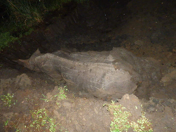
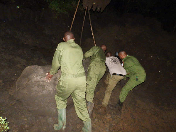
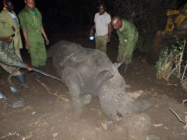
Examination and treatment: The rhino was administered with large amounts of Dextrose solution 50% I.V and i.m to re-energize it and uplift its energy levels,. However despite using ropes and straps, the rhino was unable to rise up. The body condition was quite poor with a very sharp vertebral spine an indication of starvation or illness. After a few minutes of manipulation and resuscitation the rhino succumbed.
Postmortem findings: The stomach, large and small intestines were empty due to prolonged stay without feeding, sunken eyes, sharp spinal column due to dehydration and loss of body condition. The hind legs were quite weak and flaccid a sign of nervous paralysis caused by the struggle. It had accumulation of clear peritoneal fluid and frothy lung bronchioles. The lungs bronchi were filled with muddy fluids due to partial drowning in the swamp.
The rhino died of extreme body exhaustion caused by prolonged struggle to get out of the muddy swamp. It was quite dehydrated, hypoglyceamic and the hind legs were quite flaccid and paralyzed that could not support its weight. The carcass was pulled by a tractor and buried in a deep pit nearby.
Conclusion: Its unfortunate that the rhino did not survive despite spirited efforts and teamwork by the rescue team that finally managed to get it out of the mud. We acknowledge the efforts and dedication of all the KWS and Born Free Foundation staff who participated in the exercise, we also acknowledge the efforts of the Senior Warden Meru and rhino monitoring team who mobilized resources required including the tractor borrowed from the road contractors, without their support we wouldn’t have made it.
Case #4: Treatment of an injured elephant bull
Species: African Elephant (Loxodanta africana)
Sex: Male
Age: Adult
Location: Kifuku Wildlife Conservancy
Date of intervention: 12/11/2018
History: An adult male elephant sighted at Kifuku Wildlife Conservancy with a deep penetrating gunshot wound on the left front leg that was bleeding. The elephant was quite weak and could barely move.
Immobilization, examination and Treatment: The injured elephant was darted on the thigh muscles using 18mgs of etorphine Hcl in a 1.5ml Dan-inject dart. The elephant was immobilized after about 5 minutes and went down on lateral recumbency.
The elephant had a deep penetrating wound on the posterior part of the left front leg, the wound extended caudal-anteriorly and partially breaking the radio-ulnar bone, the bones were splintered and could be felt upon probing with long forceps. The leg was also swollen due to heavy inflammation and oedema.

The wound was probed using sterile gauze swabs attached to long forceps to remove all the necrotic debri and ensure no foreign material was left inside. Thereafter it was cleaned with copious amounts of water then debrided with 10% hydrogen peroxide then cleaned and flushed with Lugol’s iodine. The wound was further treated using Opticlox® ointment applied topically and green clay applied to plug off the wound.
Other treatments were intramuscular injection of Procaine penicillin (Norocillin®) and dexamethasone to support the wound healing process. The wounds were finally sprayed using oxytetracycline spray.
Revival and prognosis: After treatment the elephant was revived from anaesthesia using 48mgs of Diprenorphine Hcl administered intravenous through the superficial ear-vein. It rose up with a lot of difficulties and walked away feeling a bit relieved. Prognosis was poor due to its inability to move and pain due to partial fracture, the security rangers were informed to monitor the progress and report to the veterinary team in case it required repeat treatment or euthanasia.
Case #5: Post-Mortem of the same elephant bull
Date of clinical intervention: 19/11/2018
History: This was a case of an adult male elephant at Kifuku Wildlife Conservancy that was treated earlier for front leg injury but later died due to a fracture and infection of the affected leg.
Postmortem examination: Postmortem examination revealed fracture of the radio-ulnar bone, there were signs of internal haemorrhage and myonecrosis on the affected part of the leg. The fracture was caused by a gun shot through the leg. Both the tusks were recovered and stored at KWS.
Cause of death: The elephant suffered from a partial fracture of the radio-ulnar bone of the left front leg, the leg became swollen and painful and the elephant developed septicaemia and died.
Case 5#: Treatment of an injured male elephant calf
Species: African Elephant (Loxodanta africana)
Sex: Male
Age: Juvenile
Location: Mpala Wildlife Conservancy
Date of clinical intervention: 14/11/2018
History: A juvenile elephant in Mpala Wildlife Conservancy sustained a a dislocation of the stifle joint of the left hind leg. The dislocation may have been caused when it got stuck in a hole or was pushed by other older elephants. The calf was in great pain and was unable to put enough weight on the affected leg. It was found with the mother and other elephants in a small family.
Immobilization, examination and treatment: The injured elephant calf was darted from a vehicle using 4mgs of etorphine Hcl. It was darted on the right thigh muscle and got immobilized after about 5 minutes and went down on lateral recumbency. The mother refused to leave the calf and the veterinary team was forced to immobilize the mother too. The mother was also darted using 17mgs of etorphine Hcl and got immobilized within 6 minutes.
The affected hind leg was swollen at the stifle joint and the joint was stiff and could not flex easily, it had no wound or any other external injury on the affected leg. It was a joint dislocation that had started healing in awkward position. The elephant calf was then treated with Vitamin B12 and Calcium (Calciject®) followed by Dexamethasone injection and multivitamin administered intramuscularly to reduce pain and enhance the recovery process.
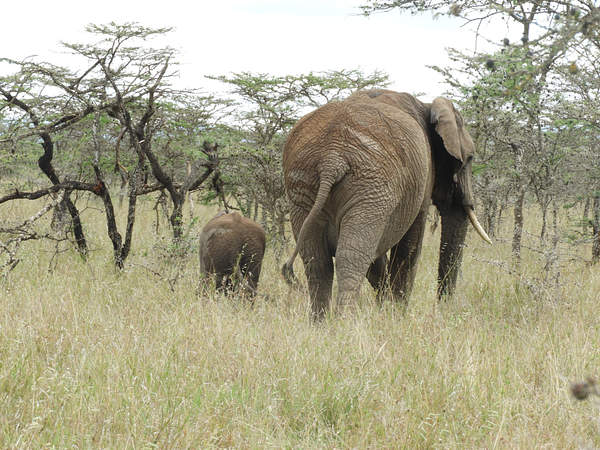
Reversal and prognosis: After treatment, the calf was revived from anaesthesia using 12mgs of diprenorphine Hcl through the superficial ear-vein. The mother was also revived and they both joined each other and walked away with the rest of the family. Prognosis was quite good as it is a young calf that with less weight and will easily recover from the joint dislocation. The calf is being closely monitored by the security team at the Mpala Conservancy who have reported good progress.
Our mobile vet initiative is in the field every day saving wild lives
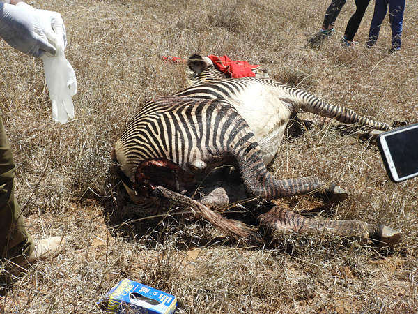
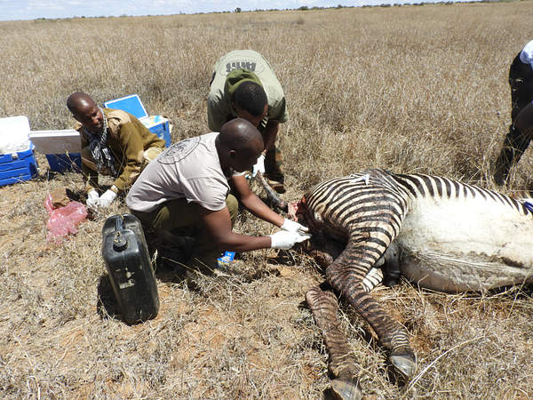
Case #6: Euthanasia and postmortem examination of a Grevy's zebra
Species: Grevy’s zebra (Equuis grevyi)
Sex: Male
Age: Adult
Location: Mpala Wildlife Conservancy
Date of clinical intervention: 15/11/2018
History: An adult male Grevy zebra that a complete compound fracture of the humerous bone of the left front leg.
Immobilization, examination and euthanasia: The injured Grevyi zebra could not move and remained in one location for quite some time. It was darted from a vehicle using 5mgs of etorphine Hcl combined with 80mgs of azaperone delivered by Dan-inject remote delivery system.

The fractured leg was examined and found to have no chances of recovery, a decision was made to euthanize the zebra to relieve it from pain and suffering and do post-mortem examination. The humerous was broken mid-shaft that could not recover, it was reported to have got fractured while running and got into a deep burrow in the field.
Case #7: Treatment of an injured female elephant
Species: African Elephant (Loxodanta africana)
Sex: Female
Age: Sub-adult
Location: Enasoit Wildlife Conservancy
Date of clinical intervention: 15/11/2018
History: A sub-adult male elephant was sighted by community scouts from Enasoit Wildlife Conservancy in Laikipia County with several wounds on the medial side of the left front leg.
Immobilization, examination and treatment: The elephant had septic wounds on the medial side of the left front leg. The affected leg was swollen and oedematous. The wounds were cleaned with lots of clean water then probed using sterile gauze swabs to remove all the necrotic debri and exudates. Thereafter the wounds were then debrided with 10% hydrogen peroxide then cleaned and flushed with tincture of iodine. It was further treated using Opticlox® ointment and green clay applied topically and oxytetracycline spray applied.
Other treatments were intramuscular injection of Procaine penicillin (Norocillin®) and dexamethasone to support the wound healing process.
Reversal and prognosis: After treatment, the elephant was revived from anaesthesia using 48mgs of Diprenorphine Hcl administered intravenous through the superficial ear-vein. It rose up successfully and walked away.
Prognosis was quite good after treatment and quick recovery was expected since the wound was higher up with minimum contamination and only affected soft tissues. The security rangers at Enasoit were informed to monitor the progress and report to the veterinary team in case it required repeat treatment.
Case #8: Treatment of a wounded male Grevy's zebra
Species: Grevy’s zebra (Equuis grevyi)
Sex: Male
Age: Adult
Location: Loisaba Wildlife Conservancy
Date of clinical intervention: 20/11/2018
History: An adult male Grevy's zebra was reported to have been attacked by a pride of lions and severely wounded on the thighs, perineum, shoulder and neck. The wounds were large and extensive and started becoming septic.
Immobilization, examination & treatment: The wounds were already septic and had developed some proud flesh with some loose skin hanging from the wound. The wounds were cleaned with lots of water, loose hanging ski trimmed off, cauterized and debrided using 10% Hydrogen peroxide and thereafter covered with povidone iodine, Opticlox® ointment then oxytetracycline spray and green clay applied to plug the wound and enhance healing process.

Further treatments using Amoxycillin (Betamox®) was administered and flunixin meglumine anti-inflammatory drug administered intramuscularly to enhance wound healing process.
Reversal and prognosis: The anaesthesia was revived using 12mgs of diprenorphine Hcl combined with 50mgs Naltrexone Hcl administered through the jugular vein. The zebra rose up after 2 minutes and ran away feeling relieved. Prognosis was favourable after treatment but the animal was quite weak and susceptible to further predation.

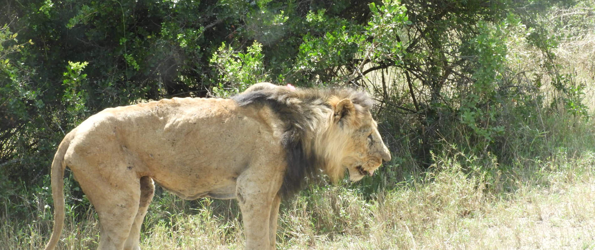
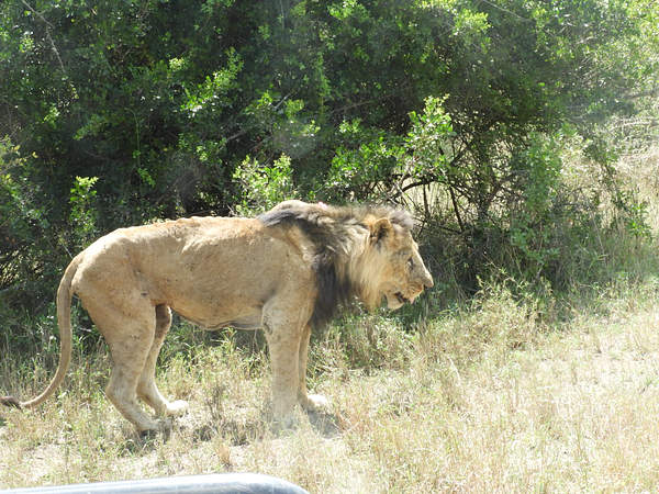
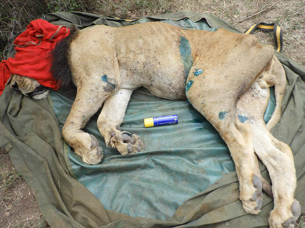
Case #9: Treatment of a sick lion
Species: Lion (Panthera leo)
Sex: Male
Age: Adult
Location: Solio Wildlife Conservancy
Date of clinical intervention: 21/11/2018
History: An adult lion in Solio Wildlife Conservancy was sighted emaciated, weak and could not hunt and feed itself. It was heavily infested with ticks on the abdomen, shoulder and neck regions. The general observation revealed the lion was in poor body condition and had no visible traumatic injury or wound on its body being weak and emaciated. It preferred lying down most of the time.
Immobilization, examination & treatment: The lion was anaesthetized using a combination of 300mgs of Ketamine Hcl and 4mgs of Medetomidine Hcl in a 3ml dart. Darting was done from a vehicle using a Dan inject® dart rifle. Anaesthesia took effect after about 10 minutes and the lion became recumbent.
The lion had lots of ticks all over the body which was sprayed adequately using Frontline® to eradicate any ectoparasites, engorged ticks were physically removed by hands. It was treated using Procaine penicillin administered intramuscularly followed by multivitamins intramuscularly.
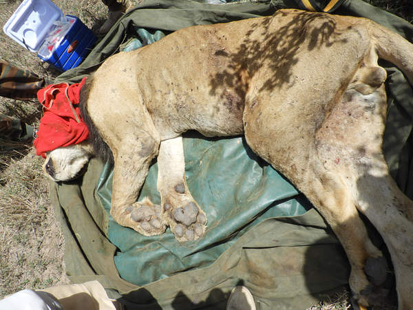
Samples collection: Biological samples including blood, tissue and ticks were collected and preserved appropriately for laboratory investigations and research.
Reversal and prognosis: After treatment anaesthesia was revived using 15mgs of atipamezole Hcl given intra-muscularly. The lion gradually recovered from anaesthesia and was up within 5 minutes. It was immediately fed on beef and it regained energy and started walking away.
The prognosis was good after treatment and removal of the ticks. It was recommended that it be supported by providing food and water until it recovers and gained energy to hunt for itself.
ACKNOWLEDGEMENT
Report by KWS Vet Dr Mijele. We acknowledge the David Sheldrick Wildlife Trust (DSWT), KWS team in Laikipia and at the veterinary department and at Mountain Conservation area and other partners for supporting the Mt. Kenya Veterinary Unit to respond and attend to many wildlife cases that required urgent veterinary attention particularly the many elephant cases that are really affected by human-elephant conflicts in the Laikipia region.